
Written by Anjani Mishra

Spinal
cord
The spinal cord is that part of central nervous system
which is caudal to the brain and contained in the vertebral canal. The cranial
end of the spinal cord is continuous with the medulla oblongata of the brain at
the level of foramen magnum of the skull.
In the different domestic animals the caudal end of
the spinal cord varies in location from the next-to-lumbar vertebra to the
middle of the sacrum.
- Ruminant- to the level of the cranial half of the 2nd sacral vertebra.
- Equine- to the level of the caudal half of the 2nd sacral vertebra.
- Porcine- between the cranial edge of the 2nd sacral vertebra and the middle of the 3rd sacral vertebra.
- Carnivore- ends very close to the junction of the 6th and 7th lumbar vertebrae.
- Aves- to the minute part of the vertebral canal within the pygostyle.
- Human- in man the ascend of the spinal cord is so great that the end of the spinal cord is approximately at the junction of the 1st and 2nd lumbar vertebra.

The spinal cord is divided into cervical, thoracic, lumbar, sacral and coccygeal or caudal parts.
- The caudal 3 cervical and cranial 2 thoracic spinal cord segments are larger in diameter and form the cervical enlargement. The enlargement is due to the increase number of nerve cells and fibers in this area which are related to the brachial plexus and the muscles of the pectoral limbs.
- The lumbar enlargement occurs in the last 3 lumbar and first 2 or 3 sacral spinal cord segments which are associated with the lumbosacral plexus and the pelvic limb.
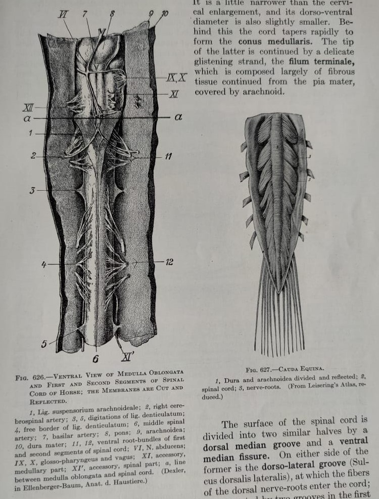
- The caudal extremity of the spinal cord tapers to a point caudal to the lumbar segments and is referred to as the conus medullaris. From the conus medullaris a slender non-nervous filament of the pia mater, the filum terminale, extends caudally in the dural sac. The filum terminale becomes incorporated in the filum of the spinal dura mater at the caudal end of the dural sac.
- The caudal portion of the spinal cord and roots of spinal nerves attached to it, because they resembles the tail of horse, and are referred to as the cauda equina.
Upon cross section of spinal cord, it can be divided into two parts;
1.
The
gray matter or gray substance
2. The white matter
1.
The gray matter- located deeply
and composed of nerve cell bodies predominantly.
- The gray matter of the spinal cord is arranged in columns which extend the entire length of the spinal cord.
- The gray matter of the spinal cord in cross section is in the form of the letter ‘H’.
- The dorsal protuberance on either side is the dorsal horn and the ventral protuberance is the ventral horn.
- The dorsal and ventral horns are the largest in the cervical and lumbosacral region of the spinal cord, because of the large number of nerve cells associated with the limb musculature.
- The central intermediate substance is the gray substance surrounding the central canal.
- The gray matter dorsal and ventral to the central canal is called the dorsal and ventral gray commissures and contain a large number of nerve fibers, particularly in the dorsal commissure.
- The central intermediate substance is continuous with the lateral intermediate substance, which is located between the dorsal and ventral gray horns.
2.
The white matter- located
superficially and composed of nerve fibers predominantly. The central canal is
the remains of the cavity of the embryonic neural tube in the spinal cord.
The white matter
is divided into three main regions by the dorsal and ventral rootlets of the
spinal nerves;
a.
Dorsal
funiculus
b.
Ventral
funiculus
c. Lateral funiculus
a.
Dorsal funiculus- the region between the dorsal median sulcus and the septum on the midline and the
dorsal rootlets and the dorsal gray horn laterally.
b. Ventral funiculus- the region between the ventral median fissure and the ventral rootlets.
c. Lateral funiculus- the region between the dorsal and ventral rootlets of the spinal nerves bordered medially by the gray matter of the dorsal, ventral and lateral horns.

Spinal cord Vs vertebral column
During early development, the segment of
the spinal cord are centered on the level off the intervertebral foramina.
Later as the vertebral column increases in length more than the spinal cord,
the nerve roots must course caudally alongside the spinal cord, as the interval
between the origin of the spinal nerve and its intervertebral foramina of exit
increases.

Facebook Veterinary group
Post a Comment