
Written By Anjani Mishra

THE AXIAL SKELETON
The axial skeleton is the branch of skeleton which deals with the bones of the head, vertebral column, ribs and sternum.
BONES OF THE HEAD

THE SKULL BONES
The skull provides a means of the protection for the
brain, the organs of special senses (sight, smell, hearing, equilibrium and
taste), the openings for the passages of air and food, and the jaws, including
the teeth for mastication. The term ‘‘cranium’’ (bones of the skull) is
sometimes referred to as consisting of those bones which lodge and protect the
brain devoid of the mandible and facial bones. According to the Nomina
Anatomica Veterinaria (NAV);1968 divides the skull in to 2 groups namely:-
·
Bones of the cranium
&
·
Bones of the face
# Formula to remember paired and single bones of skull
@ SHEVOM- Single bones
@ all the rest- paired bones
# Formula to separate cranial and
facial bones of skull
@ SOFTPIE- Cranial bones
@ all the rest- facial bones
CRANIAL BONES(paired)
Parietal Bone:-
The parietal bone is a paired structure and forms the
dorsolateral wall of the cranium with the cccipital bone caudally and the
frontal bone rostrally. It is composed of a parietal plane, temporal plane and
a nuchal plane (in the ox). Internally the grooves and ridges correspond with
the gyri and sulci of the brain. There is also an interparietal bone between
the occipital bone and the parietal bone which fuses with age.
Interparietal:-
The interparietal bones are a median bone situated
between the squamous part of the occipital bone and the parietal bone. The
bones of either side fuse with each other and with the parietal bone.
Frontal Bone:-
The frontal bone is a paired structure joined by the
interfrontal suture between the cranium and the face and enclosing the frontal
sinuses. The nasal and lacrimal bones border the frontal squama section and
form the zygomatic process laterally and part of the orbit dorsally. Lacrimal
glands are also present near the
orbit. The temporal line extends into
the external sagittal crest.
Temporal Bone :-
The temporal bone is
composed of squamous, petrosal and tympanic parts and forms the lateral wall of
the cranial cavity. It articulates with the frontal, parietal and sphenoid
bones. The squamous element joins the temporal process of the zygomatic bone to
form the zygomatic arch and forms the articulating surface of the
temporomandibular joint. An articular tubercle and mandibular fossa are
present.

CRANIAL BONES(unpaired)
Occipital Bone:-
The occipital bone forms the nuchal wall and the
foramen magnum. The pars basilaris element is the caudal base of the cranium,
although rostral to foramen magnum and joined by a cartilaginous suture to
basisphenoid bone. A nuchal crest is present and is easily palpable. The nuchal
crest is often used as a landmark for collection of cerebrospinal fluid (CSF).
There are also external occipital protuberances present which provide muscle
attachment sites for the nuchal ligament. The lateral parts form the borders of
foramen magnum. Occipital condyles are present which articulate with the atlas
to form the atlanto-occipital joint. The paracondylar process provide muscle
attachment sites for muscles of the head. The hypoglossal canal is also within
this structure.
Sphenoid Bone:-
The sphenoid bone forms the base of the neurocranium
and is composed of a body and wings. The bones are separated by cartilage which
ossifies with age. The presphenoid is rostral and has a caudal fossa which is a
hollow body with sphenoid sinuses located inside. The pterygoid processes and
oval foramen are also present in this structure.
Ethmoid Bone:-
The ethmoid bone forms part of the cranial and facial
parts of the skull and is located deep in the orbit. External lamina consist of
the roof plate, floor plate and paired orbital plates. The ethmoid bone is
separated from the cranial cavity by the cribriform plate. Numerous small
foramina exist where the olfactory nerve (CN I) passes through. The
perpendicular plate splits the ethmoid into two halves and the ethmoid
larbyrinth protrudes from the ethmoid tubes. The tubes are composed of two rows
of ethmoturbinates and air filled ethmoidal meatuses. Secondary ethmoturbinates
may also be present. Ethmoturbinates are divided into endoturbinates and
ectoturbinates. The first endoturbinate forms the dorsal nasal conchae and the
second endoturbinate froms the middle nasal conchae. The endoturbinates form 3
nasal meatuses; the dorsal nasal meatus, the middle nasal meatus and the
ventral nasal meatus.


FACIAL BONES(paired)
Maxilla:-
The maxilla forms most of the facial part of the skull, including the lateral walls of the face, nasal cavity, oral cavity and hard palate. It also forms the ventral nasal conchae and articulates with all of the facial bones as it is the largest bone of the face.
Premaxilla:-
The premaxilla (incisive) bones are the most rostral
bone of the face and are each composed of a body, a palatine process and a
nasal process. The bone is narrower and more pointed rostrally in case of ox.
Lacrimal:-
The lacrimal bone forms part of the lateral wall of
the face and orbit and is situated near the medial canthus. It articulates with
the frontal bone, zygomatic bone and maxilla. It also articulates with the
nasal bone in ruminants and the horse and articulates with the palatine bone in
carnivores. It is composed of an orbital and facial part separated by supra-
and infraorbital margins. The nasolacrimal duct is present by the margin of the
orbital surface. The ventral oblique muscle attaches caudal to the margin of
the orbital surface. The nasal surface forms the boundaries of the maxillary
and frontal sinuses.
Malar/Zygomatic:-
The molar (zygomatic) bone is in the caudolateral area
of the face and forms the ventral border of the orbital cavity.
Pterygoid:-
The pterygoid bone is a paired structure bordered by
the palatine and sphenoid bones. It forms the dorsal and lateral walls of the
nasopharyngeal cavity. The pterygoid hamulus is formed by the pterygoid bone.
Nasal:-
The nasal bone is a paired structure and forms the
roof of the nasal cavity. Dorsal nasal conchae attach to the ethmoidal crest on
the internal surface. A rostral suture forms the apex and between the nasal and
incisive bones is the naso-incisive notch.
Palatine:-
The palatine bone is a paired structure between the maxilla, sphenoid and pterygoid bones. It is composed of horizontal plate(forms part of the hard palate) on which nasal crest is present. The palatine sinus is present on horizontal plate.
Turbinate bones:-
These are delicate, scroll-like bones(four in number) which are attached to the lateral wall of the nasal cavity. Each bone is composed of a very thin lamina, cribriform in many places and covered on both sides with mucous membrane in the fresh state. They are arranged in two pairs, dorsal and ventral.

FACIAL BONES(unpaired)
Vomer:-
The vomer is unpaired and extends from the choanae of
the palatine bone to the floor of the nasal cavity. It attaches to the median
nuchal crest and has a septal sulcus which surrounds nasal cavity.
Mandible:-
The mandible can be divided into the body and the
ramus. The body of the mandible supports the incisor teeth (rostrally) and
cheek teeth (caudally). The section of the body which does not support any
teeth is called the inter-alveolar margin or diastema. The mandible also
contains the mandibular canal and the mental foramen. The facial notch is on
the ventral surface where the parotid duct (in herbivores) and facial vessels
run. The ramus extends from the caudal end of the body dorsally towards the
zygomatic arch. The condylar process
articulates with the mandibular process of the skull.

Here, we see the bones of skull are flat bones,
developed in membrane and the bones of the cranial base may be classed as
irregular and are developed in cartilage . Only two form are permanent movable
joints with other part of skull .The mandible form synovial joint with the
temporal bone, and the hyoid bone is attached to the latter by bar of
cartilage. The immovable joint located between most of the bones of skull are
termed ‘suture’. But these suture disappear by osseous fusion on increasing age
of animal. The skull present numerous foramina as mention below: - infraorbital,
mental, mandibular, supraorbital, etc. through which cranial nerves and blood
vessels enter and exit.
Hyoid Bone:-
· The hyoid bone(Os
hyoidiun) is situated chiefly between the vertical parts of the rami of the
mandible, but its upper part extends somewhat further back.
· It is attached to
the petrous temporal bones by rods of cartilage, and supports the root of the
tongue, the pharynx, and the larynx.
It consists of a
body, a lingual process, and the four pairs of cornua.
Body:
· The body
is a short transverse bar, compressed dorso-ventrally.
· The dorsal surface
is concave and smooth in its middle, and presents a convex facet or tubercle at
each end for articulation with the small cornu.
· The ventral
surface is flattened and is slightly roughened for muscular attachment.
· The anterior
border carries the lingual process medially.
· The posterior
border is concave and smooth in its middle and carries the thyroid cornu on either
side.
Lingual
process:
· It has a short
tuberous lingual process.
· The lingual
process projects forward medially from the body, and is embedded in the root of the tongue.
Cornua:
A.
The thyroid cornua
or thyroids
· They extend
backward and upward from the lateral parts of the body.
· They are
compressed laterally(except at their junction with the body), and the posterior
end has a short cartilaginous prolongation which is connected with the anterior
cornu of the thyroid cartilage of larynx.
B.
The small cornua
or keratohyoids
· They are short rods which are directed upward
and forward from either end of the body.
· Each of which is somewhat constricted in its
middle part and has a slightly enlarged ends.
· The ventral end has a small concave facet
which articulates with the body.
· The dorsal end articulates with the great
cornu(horse), or with the middle cornu when present.
C.
The middle cornua
or epihyoids
· These are small,
wedge-shaped pieces or nodules interposed between the small and great cornua.
· They are usually
transitory, and unite with the great cornua in the adult.
· The middle cornua
are almost as large as the small cornua.
D.
The great cornua or stylohyoids
· They are the
largest parts of the bone.
· They are directed
dorsally and backward, and are connected above with the base of the petrous
temporal bone.
· Each of which is a
thin plate, which is slightly curved in its length, so that the lateral surface
is concave and the medial surface is convex. Both surfaces are smooth.
· The border are
thin. The dorsal extremity is large and forms two angles; the articular angle
is connected by a rod of cartilage with the hyoid process of the petrous
temporal bone whereas the muscular angle is somewhat thickened and rough for
muscular attachment.
· The ventral extremity
is small, and articulates with the small or middle cornu.


THE VERTEBRAL COLUMN
- It is the
part of axial skeleton.
- It is an
articulated structure of vertebra, cartilage and ligaments that gives
passage to the spinal cord and its covering(meninges).
- Commonly
called spine of the body.
- It comprises of five regions:
|
Vertebra |
Region |
|
|
Definition:
Vertebral column is the fundamental part of axial skeleton consists of chain of median, unpaired, and irregular bones which extends from the occipital bone to the end of the tail.
The vertebral column is subdivided for
description into 5 regions, which are named A/to the part of the body in which
the vertebrae are situated. Thus the vertebrae are designated as cervical,
thoracic, lumbar, sacral and coccygeal(caudal) vertebrae. The number of
vertebrae in a given species is fairly constant in each region except the last,
so that the vertebral formula may be expressed(for example in case of ox) as
follows;
OX- C7, T13 , L6, S5,
Cy18-20
Vertebral formula- express the total
number of bones present
in the vertebral column.
Vertebral formula of domestic animals and fowl:
|
Species
|
Cervical |
Thoracic |
Lumbar |
Sacral |
Coccygeal |
Total(Av.) |
|
Ox |
C7 |
T13 |
L6 |
S5 |
Cy18-20 |
50 |
|
Horse |
C7 |
T18 |
L6 |
S5 |
Cy15-21 |
54 |
|
Sheep/Goat |
C7 |
T13 |
L6-7 |
S4 |
Cy16-18 |
48 |
|
Dog |
C7 |
T13 |
L7 |
S3 |
Cy20-23 |
51 |
|
Pig |
C7 |
T14-15 |
L6-7 |
S4 |
Cy20-23 |
53 |
|
Fowl |
C14 |
T7 |
(L+S)14 |
Cy6 |
41 |
|
TYPICAL VERTEBRA:
The vertebrae in a given region have characteristics by which they may be distinguished from those of other regions, and individual vertebra have characteristics which are more or less clearly recognizable. All typical vertebrae have a common plan of structure, which must first be understood. A vertebra consists of a body, arch and processes.
- Mass of bone
on which vertebra is built.
- It is
cylindrical, solid and rod like.
- The dorsal
surface of the body forms the floor of neural (spinal) canal.
- The ventral
surface presents ill developed spines called infraspinatus process, for
muscular attachment.
- The centrum
is convex anteriorly and concave posteriorly.
- The vertebra are articulated to each other both at the centrum and oblique process.
Arch:
- The neural arch consists of lamina and the pedicle, together. (Dorsally, from either side of the body spring plates of bones called pedicles which form the wall of the neural ring. They unite above to form the roof and are called laminae).
- The arches of
the opposite sides unite together to complete the neural ring.
- Series of
neural rings with intervertebral membrane constitute the neural canal,
which lodges the spinal cord.
- The pedicles bear notches at their cranial and caudal aspects, which form a circular opening together with preceding vertebra called intervertebral foramen, through which the spinal nerves and vessels pass.
Processes:
A) Spinous processes:
a) Supraspinous
process/Neural Spine/ Dorsal spine:
·
It
is a single process project dorsally form the middle of the arch, where the two
laminae meet.
·
It
varies greatly in form, size and direction in different vertebrae.
· It furnishes attachment to muscles and ligaments.
b) Infraspinous
process:
- Its is on the
ventral part of the centrum.
- Some vertebrae also have a ventral tubercle, ventral crest or a hemal arch.
B)
Oblique/Articular Processes:
- The vertebral
segments articulate with each other, both at centrum and at oblique
process.
- The oblique
processes are four in numbers, two being placed anteriorly and two
posteriorly.
- They bear a
small articular part.
- The anterior
oblique process faces above and articulates with the posterior oblique
process of the preceding vertebra, which look ventrally.
- They are called oblique process due to their placement.
C)
Transverse processes:
- Are two in
number and project laterally from the sides of the arch or from the
junction of the arch and body.
- In the
cervical region the transverse processes of the 3rd to 6th
cervical vertebrae present a cranial and caudal portion.
- The transverse processes are pierced by the transverse foramen(foramen transversarium) at their base.
D) Mamillary processes:
- Are found in most animals on the caudal thoracic and cranial lumbar vertebrae between the transverse and cranial articular processes or on the latter.
E) Accessory processes:
- When present, are situated between the transverse and caudal articular processes.
THE CERVICAL VERTEBRA
- The vertebral
segments of neck (cervical) region are called cervical vertebra and are
seven in number.
- Placed
serially one behind the other, being designated as first (atlas), second
(axis), third, fourth, fifth, sixth and seventh cervical vertebra.
- They have
long cylindrical centrum. They decrease in length from before backwards,
while their width increases.
- The oblique
processes are well developed and form diarthrodial joints.
- The first cervical vertebra(atlas) is completely an atypical where as the second cervical vertebra(axis) is partially an atypical or typical. Rest of the vertebrae of this segment are typical.
Atlas
· It
widely differs from the rest of vertebrae, hence, it is called an atypical
vertebra.
· In domestic animals head is suspended form atlas where as in human, atlas carries the head.
- It presents a ring and two large plates of bone, termed wing or alae.
Ring:
· Ring
is more or less rounded.
· It
has 4 surfaces: anterior, posterior, dorsal and ventral.
· The
dorsal surface presents a raised eminence called dorsal tubercle, which
represents supraspinous process of other vertebra.
· On
the ventral surface there is an obtuse eminence called ventral tubercle.
- The anterior surface present two foramen; vertebral foramen (antero-internal) and the foramen alare (antero-external) connected by a short furrow.
· On the posterior surface there is a
smooth articular surface called fovea dentis, which lodges odontoid process of
the axis.
· The ring anteriorly presents on
either side a deep articular cavity which receive the occipital condyle of
skull.
- The ring posteriorly presents
two articular surface (fovea dentis and lateral articular surface) for
articulation with the anterior surface odontoid process and articular
surface) of axis.

Wing
or Alae:
· The
wing represents transverse process of the typical vertebra.
· It
presents dorsal and ventral surfaces.
· On
the dorsal surface of the wing just behind the foramen alare (1st)
and vertebral foramen (2nd), there are the external openings of the
third and fourth foramina.
· On
the ventral surface at the junction of the ring, there is a deep fossa
atlantis.
· Foramen transversarium is absent.
Axis
· It
is longest of the vertebrae and partly atypical.
· On
the anterior part of the body, there is a projection called odontoid process
(dens), which is dorsally deeply concave and rough and ventrally articular,
smooth and convex.
· On
the either side of the odontoid process, there are extensive, nearly flat
articular surface, which may be considered as modified anterior oblique
process. They blend with the ventral surface of the odontoid process. The
entire surface thus, articulates with the fovea dentis of the atlas.
· The
intervertebral foramen is present a little behind the anterior notch, which is
circular.
· The
infraspinous process is in the form of median ridge and the supraspinous
process projects a little infront over the canal.
· Posterior
oblique process placed posterolateral over the canal.
· The foramen transversarium is either small or absent.

The
third, foUrth and fifth cervical vertebra
·
These
vertebrae are similar with typical vertebrae.
·
In
serial placement the bodies tend to become shorter and wider form front to
backwards.
·
The
supraspinous process is short and tuberous centrally in the third, then
gradually increases in height and length upto the seventh with forward
inclination.
·
Ventrally,
projects a well defined infraspionus process with increseas in length and angle
of descend.
·
The
oblique processes are slightly convex anteriorly and concave posteriorly, the
size of which increases with the receding number.
·
The
oblique processes are slightly convex anteriorly and concave posteriorly, the
size of which increases with the receding number.
·
The
transervse processes are divided into upper tubercular portion (project at
right angles) and lower plate like portion directed outwards, downwards and
forwards.
·
At
the base of tubercular transverse process, the foramen transversarium is
present. Series of these foramina continue to form canalis transversarious.
The centrum is convex anteriorly and concave and deep posteriorly.

THE sixth cervical vertebra
· It
is a typical vertebra.
· The
supraspinous process is well developed, while the infraspinous process is
absent.
· The
oblique process is larger.
· The transverse process is divided and the lower part is modified to form ventral branch of transverse process and laterally it forms lateral branch of transverse process, and is pierced at the base by the foramen transversarium, which is very large (largest in the series).

THE seventh cervical vertebra
· It is a typical vertebra.· It bears the characters of both cervical and thoracic segments.
· The supraspinous process is highest in the series.
· Infraspinous process is represented by two tubercles.
· The foramen transversarium is absent.
· The transverse process is undivided.
· The neural ring is very large.
THE THORACIC OR DORSAL VERTEBRAE
· There
are 13 thoracic vertebrae in this region.
· The
vertebrae of this region are characterized by the presence of articular surface
on the bodies for the heads of the ribs and on the transverse process for the
tubercles of the ribs; and by the excessive development of supraspinous
process.
· The
supraspinous process is well developed and highest.
· The
infraspinous process is in the form of thin ridge.
· The
oblique process is small. The anterior oblique process is slightly convex and
placed on the dorsal part, directed upward. The posterior oblique process are
slightly concave and placed at the base of dorsal spine, face downwards.
· The
transverse process are short, thick and single.
· Each
transverse process bears a tubercular facet on its ventral aspect for
articulation with the tubercle of rib. They bear a thick rounded mamillary
process on the dorsal aspect, which may be absent or ill developed in last few
segments.
· The
bodies are short, distinctly constricted in the middle and are expanded at the extremities.
· On
the either side at either end of the articular extremities of the centrum,
there is a concave articular surface, termed as costal facet. Each is a
demi-facet as it articulates with half the part of the head of rib.
· The
vertebral notches are shallower and smaller but anterior notches are deeper.
· The
arch is perforated by an additional intervertebral foramen on either side, in
the posterior segments.
· Spine of the first is pointed backwards. This backwards inclination increase up to the tenth and then decrease.
· The summits of the seventh to tenth are distinctly bifid.

The first thoracic vertebra
· It shares the characters of both the thoracic and cervical vertebrae.
· The
costal and tubular facets are the largest in the series so also the transverse
process
· The
supraspinous process suddenly rises in height
· The anterior oblique process resemble those of cervical region.

The LAst thoracic vertebra
· The
body is oval and bears no posterior pair of costal facet.
· The supraspionus process is vertical of slightly incline forward.
The lumbar vertebrae
· There
are six lumbar vertebra and form the skeleton of loins.
· Excessive
development of transverse process is a special character.
· Each
transverse process is a long plate of bone, the transverse processes are separated by intertransverse space. The length of first transverse process is
the shortest. It gradually increases up to 4th & 5th.
The 6th is slightly shorter than the 5th.
· The
supraspinous process is broad and flattened.
· The
oblique process is well developed. The anterior oblique process is strongly curved, facing inwards, while the posterior ones are correspondingly convex.
· The
mammillary process are fused with the anterior oblique processes , which are thick and tuberous.
· The
bodies of lumbar vertebra are constricted in the middle, expanded at the extremities and resemble as thoracic vertebrae.
· The
neural canal is uniform up to 3rd and increase in length and height
up to the last.
· The
posterior notches are larger than the anterior ones.
· The intervertebral foramen frequently doubles in the anterior few segments.

THE first lumbar vertebra
· Body
is small and cylindrical.
· Articulates
anteriorly with the last thoracic segment.
· Space between the two anterior processes is the shortest in the series, so also the intertransverse space.
THE sixth lumbar vertebra
· Body
is compressed form above downwards.
· Neural
ring is very wide.
· Oblique
processes are placed wide apart.
· Supraspinous process is smallest in series.
THE Sacrum
The sacrum
consists of five sacral bones fused
together to form a single bone. It forms the region of croup.
Direction: it is
horizontal in direction.
Location: Located between
lumbar vertebrae anteriorly and coccygeal vertebrae posteriorly.
Relation : it
articulates with the ilium, anteriorly with last lumber and posteriorly with
the first coccygeal vertebra.
Shape: it is aeroplane shape.


Composition: it present 2 surfaces, 2 borders, a base and an
apex.
Surface:
Dorsal
surface:
·
Presents
centrally five sacral spines, which are fused together to form the medial
sacral crest.
·
On
other side of the base of the median sacral crest are the lateral sacral crest,
which are the fusion of the oblique processes.
·
There
are 4 pairs of dorsal sacral foramina, of which the 1st & 2nd
pairs are located internal to lateral sacral crest, and the remaining are
external to the lateral sacral crest.
·
Neural
rings are triangular, anterior larger and posterior smaller.
Ventral
Surface:
·
It
forms the roof of the pelvic cavity and is concave from side to side and from
before backwards.
· It presents 4 pairs of ventral sacral foramina, and 4 transverse lines and one central median longitudinal furrow (sulcus vasculosus).
Borders:
The two
lateral borders are thin, sharp, concave and irregular.
Base:
·
It
is the anterior extremity of the first sacral vertebra.
·
It
consists of body and two alae or wings.
·
The
body is located centrally and is concave.
· The wings are anteriorly concave and articulate with ilium by auricular facet.
Apex:
·
Apex
is the posterior end of 5th sacral vertebra and is slightly wider
than the 4th sacral vertebra.
·
It
has centrally a small triangular neural ring.
· On either side of the centrum of the last segment are backward projections, which represent the transverse process of the last sacral vertebra.
Comparison
with:
A) Sacrum of Horse: (S5)
·
Supraspinous
process is distinct.
·
Four
dorsal sacral foramina are on either side of the base of spines.
·
The
lateral sacral crest is suppressed.
·
Lines
transversae are less distinct.
·
The
ventral sacral foramina are smaller than that of ox.
· The auricular surface is elongated, oval and large.
B) Sacrum of Dog: (S3)
·
Median
sacral crests are in the form of two tubercles.
·
Lateral
sacral crests are two in number.
·
The
wings are high and auricular surfaces are directed outwards.
· The ventral foramina are two in number.
C) Sacrum of Pig: (S4)
·
Supraspinous
process is ill developed.
·
Body
is less curved.
· Cranial articular processes are well developed.
D) Sacrum of Fowl:
(L+S14)
·
Lumbar
and sacral vertebrae are fused to form a single synsacrum.
· This bone remains articulated with the ilia along their lateral borders.
THE COCCYGEAL VERTEBRA
·
The
coccygeal or tail region consists of 15-20 segments.
·
In
the first three of four, all the characteristics features of a true vertebra
are found.
·
The
first coccygeal vertebra may be fused with sacrum.
·
The
first five have complete neural arches, supraspinous processes and large
transverse processes. Anterior oblique processes are very distinct, but don't
form ant articulation.
·
The
first 12 present two small processes on the ventral surface of the bodies, the haemal processes, which pass the middle
coccygeal artery.
·
Oblique
processes disappear from the 7th or 8th.

Comparison of different vertebrae
with:
A) Horse:
C7
T18 L6 S5 Cy15-21
·
Body
of cervical vertebra is longer.
·
Wing
of the atlas are pierced posteriorly by foramen transversarium.
·
Mamillary
processes are in few caudal segments of thoracic vertebrae which are more
developed.
·
Coccygeal
vertebrae are shorter and ill developed.
B) Dog:
C7
T13 L7 S3 Cy20-23
·
Foramen
transversarium is present in the atlas at its posterior aspect.
·
In
lumbar vertebrae, the spinous processes are comparatively short, broad
ventrally and narrow dorsally.
· Coccygeal vertebrae are better developed.
C) Pig:
C7
T14-15 L6-7 S4 Cy20-23
·
Cervical
vertebrae are short and bodies are wide.
·
Thoracic
vertebrae are comparatively long.
·
Spinous
processes of cervical vertebrae are short but large in axis.
· First few coccygeal vertebrae present well-developed articular processes.
D) Fowl:
C14
T7 L+S14 Cy6
·
In
general, the vertebral column is rigid & stiff due to fusion of some
vertebral segments.
·
Atlas
is in the form of ring, presents only one articular facet for single occipital
condyle.
·
Cervical
vertebrae have styloid projections from transverse processes.
· Last few caudal vertebrae fuse to form a pointed bony projection, known as pygostyle.
THE RibS
It presents a shaft and 2 extremities.
Shaft:
It is elongated and curved more at the upper part, while it is twisted and turned inwards at the lower part.
It
presents 2 surfaces and 2 borders.
Surfaces:
·
The lateral surface is convex and
is market by a wide groove on its anterior part.
·
The medial surface is concave,
smooth and is covered by pleura.
Borders :
· Anterior border is
thick and concave.
- Posterior border is convex and
presents costal groove.
Extremities:
Proximal extremity:
·
Consists of head, neck and
tubercle.
·
Head (capitulum) is rounded and has
a hemispherical articular surface, which is divided into two convex facets,
anterior & posterior by a groove (sulcus capituli). It articulates with the
costal facet placed on the body of thoracic vertebra.
·
Neck (collum costae) is a long
constriction below the head and separates the head from tubercle.
·
The tubercle (tuberculum costae)
bears a concave facet for articulation with tubercular facet of the thoracic
vertebrae.
Distal/Sternal Extremity
- It is either articular or is prolonged by a costal cartilage.
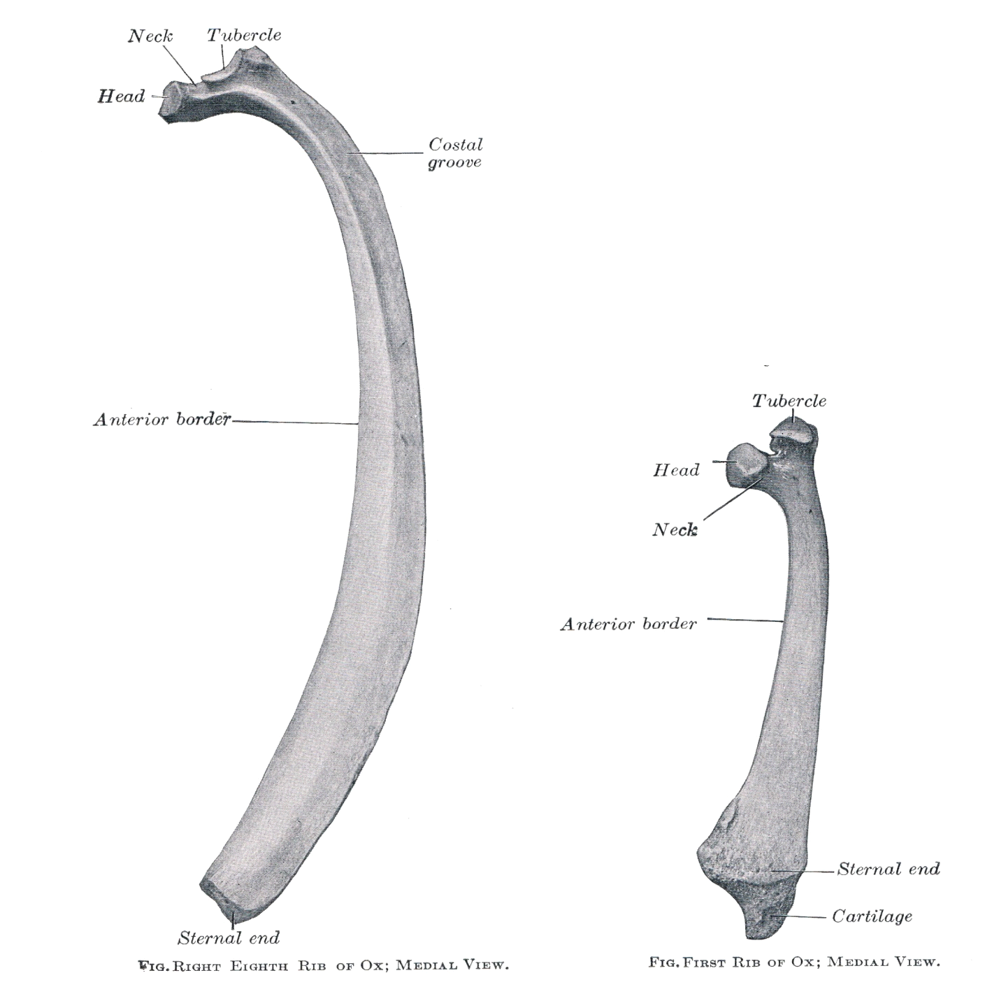
Characters of Ribs in Serial placement:
·
The first rib is shortest, thickest
and last curved. It body widens greatly towards the distal end. The costal
groove is absent.
·
The 8th, 9th
& 10th ribs are generally the longest and the widest.
·
The head and tubercle decrease in
size from the first to last ribs.
·
The curvature of the ribs increases
from the first to last.
·
The last rib is slender and curved.
The head and the tubercle of this rib are smallest and fuse with each other and
hence neck is absent.
·
The distal extremity is thin and
prolonged by the costal cartilage.
THE COSTAL CARTILAGES
·
It is the prolongation of rib,
which helps to complete the lateral walls of the thorax.
·
Each is a cylindrical piece of
cartilage, slightly compressed from sides to sides.
· It is rounded and smooth on its borders and surfaces.
Comparison with:
A) Ribs of Horse:
·
18 pairs of ribs, eight sternal and
ten asternal.
·
11th is longest and 6th
is the widest rib.
· Costal groove is distinct on the 4th to 8th ribs and absent on the first.
B) Ribs of Dog:
·
13 pairs of ribs, nine sternal and
four asternal.
·
The first eight or nine increase in
width at their distal extremity.
·
The last three of four ribs
articulate with anterior costal facets, the posterior being rudimentary.
· First rib is very long and highly curved.
C) Ribs of Pig:
·
14-15 pairs of ribs, 7 sternal and
7-8 asternal.
·
The first rib has prismatic body.
· The last rib is usually small and floating
D) Ribs of Fowl:
·
7 pairs of ribs.
·
Each rib has a dorsal 7 ventral
segment.
·
First two ribs don’t extend up to
sternum.
- Each of the dorsal segments of 2nd-6th ribs processes a caudal extension, called uncinate process, which supports the thoracic cage in a better way of overlapping the succiding rib.
The sternum(Breast bone)
·
It is centrally placed, segmented,
osteo-cartilagenous structure, forming the floor of the thoracic cavity.
·
It is held in suspension by the
costal cartilage.
·
In the adult animals, it consists
of seven segments (sternebrae), which are elongated from before backwards.
·
Sternum is compressed laterally in
the front, and dorso-ventrally behind.
·
It is oblique
direction pointing downwards and backwards.


It presents 2 surfaces, 2 borders and 2 extremities.
Surface:
Dorsal surface
·
It is flat and
rough, widens from before backwards and becomes much constricted behind the
last pair of costal facets.
Ventral surfaces :
·
It is strongly
convex anteriorly and is flat and depressed behind. It is market by a faint
median longitudinal ridge, which divides the surface into two equal halves.
·
Ventral crest is
absent.
Borders:
2 lateral borders
Lateral borders:
·
Divide the dorsal
and ventral surface. Each border separates anteriorly into dorsal and ventral
divisions to enclose 7 sternal facets.
Extremities
Anterior extremity
·
It is cranial end of the 1st
strnebra and forms the presternum or manubrium sterni.
·
It is compressed laterally and is
placed at an angle in front of the 2nd sternebra.
·
It bears a facet for articulation
with the first costal cartilage on either side.
Posterior extremity:
·
It is prolonged by
the xiphoid cartilage. It is concave above and convex below.
Comparison
with
Sternum of horse:
·
Fusion of seven sternebrae.
·
Boat shaped, compressed laterally
towards the cranial end and dorso-ventrally at the caudal end.
·
Lateral surfaces are convex.
·
Ventrally it presents a prominent
crest.
·
Xiphoid cartilage is flat and
rounded.
Sternum of dog:
·
Formed by 8 sternal segments, which
generally don’t fuse with each other. Many fuse at very old age.
·
First sternebra is longest.
·
Cranial end is blunt and the
xiphoid cartilage is narrow.
Sternum of Pig:
·
Six sternebrae has fused to form
this bone
·
Bone is dorso-ventrally compressed.
·
Xiphoid process is long and xiphoid
cartilage is small.
Sternum of Fowl:
·
It is large irregular plate of bone
composed of cranial mass.
·
Cranial mass
has:
o
Cranial projection : rostrum
o
Caudal extension: metasterum and
lateral process.
o
Dorsal surface is concave.
o
Ventrally presents a plate like
bone, which is extended up to the metasternum known as sternal crest or keel or crania.
·
The rostrum presents tow facets for
coracoids.
· The lateral processes are two in number.
· The cranial end is flat and the caudal one is divided into a dorsal and a ventral part.
mishravetanatomy@gmail.com
Facebook Veterinary group link - https://www.facebook.com/groups/1287264324797711/
Twitter - @MishraVet
Facebook - Anjani Mishra
https://uii.io/wgRR76 best video to understand anatomy
ReplyDeletePost a Comment