
Bones of Appendicular skeleton
General plan
Written By Anjani Mishra
Appendicular skeleton: is the fundamental part of the skeleton which deals with the form and structure of the bones of the limbs, i.e. forelimb and hindlimb.
Fore
limb/Thoracic limb (Four Segments)
|
Hind
limb/Pelvic limb (Four Segments)
|
Thoracic Girdle/Shoulder/Pectoral Girdle
Scapula,
Coracoid, and Clavicle
Arm (upper arm)
Humerus
Forearm
Radius/Ulna
Manus/ Forepaw
Carpus (wrist)
– Carpal bones
Metacarpus –
Metacarpal bones
Digit/digits –
Phalanges and Sesamoid bones
|
Pelvic girdle/ Hip bone/Ossa Coxarum
Ilium,
Ischium, and Pubis
Thigh:
Femur
Leg/Crus
Tibia/Fibula
Pes/ Hindpaw
Tarsus(hock/ankle)
– Tarsal bones
Metatarsus –
Metatarsal bones
Digit/digits –
Phalanges and Sesamoid
bones |
The forelimb (ox)
- Forelimb is attached to the thorax by means of muscles of the thoracic girdle.
- It is divided into four segments in each leg.
The shoulder girdle – consists of;
Scapula (2)
Coracoid (2)
Clavicle (2)- may be absent or if present, it is embedded in the brachiocephalicus muscle in the form of fibrous intersection.
The arm – consists of;
Humerus (2)
The forearm – consists of;
Radius/ulna (2)
The manus – consists of;
Carpal bones (12)
- Metacarpus – consists of;
Metacarpal bones (6)
- Digits (8) – consists of;
Phalanges (12)
Sesamoid (12)

THORACIC/ SHOULDER GIRDLE/ PECTORL GIRDLE
Shape: Scapula is a flat, more regularly triangular bone than horse.
Location: It is located on the cranial part against the lateral wall of the thorax,
being gently curved. Its long axis extends obliquely from the fourth thoracic spine to the sternal/ventral end of the first rib.
Direction: It is directed downward and forward.
Relation/Articulation: It articulates with the humerus below forming a shoulder joint and medially
attached with the thorax by means of muscles.


Composition:
It consists of 2 surfaces, 3
borders and 3 angles.
Surfaces: The scapula is wider above than below. The two surfaces are:
- Lateral Surface
- Medial Surface
Lateral Surface:
- It is divided into two unequal areas (1:3) by the spine of the scapula.
- The area in front or superior of the spine forms only one-fourth part and is termed as supraspinatus fossa and the area behind or inferior to the spine forms three-fourth of the dorsum and is termed as infraspinatus fossa.
- The supraspinatus fossa lodges supraspinatus muscles where as infraspinatus fossa lodges the infraspinatus muscles. The infraspinatus fossa also bears roughened lines to which the teres minor muscle is attached.
- The spine of scapula is wavy in outline. It marks a backward bend to about its middle and forward bend below. The free margin of spine is thickened in the middle for the attachment of the tarpezius muscle. The spine becomes more prominent below and is prolonged by a pointed projection, the acromion process which gives origin to the acromial part of the deltoid muscle.
Medial Surface/Costal
Surface:
- Consist of a shallow subscapular fossa in the middle, which lodges(origin) the subscapularis muscle.
- At the upper part of this surface, cranially, there is a rough triangular area for the attachment(insertion) of serratus cervicis muscle.
- A rough muscular line (at the caudal angle of scapula) posteriorly serves for the insertion of serratus thoracic muscle.
Borders:
Three borders: Anterior, Posterior & Vertebral.
Anterior
border: Thin, sharp and convex in the dorsal two third and thick,
rounded and concave for the remainder of its extent.
Posterior
border: Thick and slightly concavo-convex.
Nutrient foramen is on lower-third of this border.
Vertebral
border: Thick and pitted for the reception of scapular cartilage, which posteriorly forms a rounded projection.
Angles:
Three angles: Anterior/Cervical angle, Posterior/Dorsal angle & Inferior/Articular
angle.
Anterior
angle: Thin and formed by the vertebral and the anterior border.
Posterior
angle: Thick and tuberous and is formed by the vertebral and posterior
borders.
Inferior angle:
·
It is attached to the rest of the bone by a
constriction, the neck of the
scapula.
·
It is composed of a glenoid cavity and a tuber
scapula.
· The glenoid
cavity is shallow, nearly circular, articular depression and meets the head
of the humerus to form the shoulder joint.
·
The rim of the cavity presents and undeveloped glenoid notch on its lateral aspect.
The tuber scapula is
a rough eminence placed in front of the glenoid cavity and gives origin to the
tendon of the biceps brachii muscle. A short, rounded coracoid process projects from the medial side of the tuber scapula and
gives origin to the coraco-brachialis
muscle.
Spices difference of scapula:
a) Scapula of horse
- The spine is placed a little further backwards form the anterior border.
- Subscapular fossa is deeper.
- The tuber scapulae and glenoid cavity are placed further apart.
- Glenoid notch is deep and distinct.
- Acromion process is absent

b) scapula of Dog
- Spine is placed in the middle and divides the lateral surface in two equal fossae
- Acromion process is short, blunt and overhangs the glenoid cavity.
- The subscapular fossa is very shallow.
- The anterior border is thin and convex. The posterior border is thick and nearly straight. The ventral border is convex.
- Tuber scapulae is blunt.
- Glenoid cavity is prolonged forwards under the tuber scapulae.
- Small coracoid process is present.
- (Note:- the shoulder girdle has three bones- the scapula, coracoid and the clavicle. The clavicle is embedded in the brachio-cephalicus muscle in front of the scapulo-humeral articulation. Clavicle is thin, small and irregular triangular, bony or cartilaginous plate. It doesn't articulate with the skeleton.)

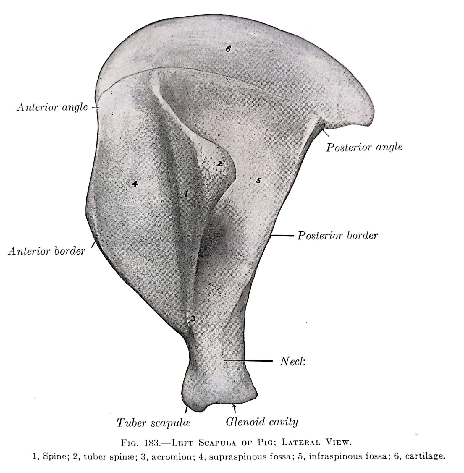
c) Scapula of Pig
- The ratio between supraspionus fossa and infraspinous fossa is 1:0.8
- Spine is wide and further directed backward.
- Acromion process is rudimentary.
- Glenoid notch is absent.

d) Scapula of goat
- Relatively smaller but more triangular and angular than ox.
- Subscapular fossa is more extensive, that is triangular in shape.

e) Scapula of Rabbit
- Spine is situated more cranially.
- Anterior angle is blunt.
- Coracoid process is developed.
- Metacromion process is present.

f) Scapula of fowl:
- It is elongated, narrow, thin slightly curved bone and situated backward parallel to the vertebral column reaching almost the pelvis.
- The anterior extremity is articular and meets the coracoid and the humerus while the posterior end is free and non-articular.
Pectoral Girdle of fowl

The
pectoral girdle are composed of three pairs of bones which support the wings.
They are formed by the fused clavicles (called furcula), the coracoids and the
scapulae.
The
most robust bone of the pectoral girdle is the coracoid, which is directed
ventrally and caudally to articulate with the sternum at the coracoidal sulcus.
The coracoids are hollow, being invaded by the clavicular air sacs.
The
clavicles are slender rod-like bones
which are ventrally fused into a flattened plate, the hypocleidium (furcular
facet), which is connected to the carinal apex of the sternum by a ligament,
the hypocleidial ligament, which also represents the raphe of the mojor flight
muscle, the pectoralis.
The long, flat scapula extends caudally, paralleling
the vertebral column; it is slightly thicker at its proximal end, and near the
acromion process there is a pneumatic foramen. The scapula articulates
cranially with the coracoid and furculum and participates with the former in
the formation of the glenoid fossa, the articular fossa for the head of the
humerus.
The three bones scapula, coracoid and furculum- come together dorsally
leaving a triosseal canal (foramen triosseum) through which the tendon of the
supracoracoideus muscle passes to insert on the humerus as an important part of
the flight mechanism; it acts to elevate the humerus and the wing.


If you have any questions you can ask me on :
mishravetanatomy@gmail.com
Facebook - Anjani Mishra
Website: mishravetanatomy.blogspot.com
sir can you expalain more about scapula of fowl including the str of spine,tuber scapulae
ReplyDelete,glenoid cavity,bordrs etc...
Post a Comment