
Written by Anjani Mishra

STOMACH
The ruminant stomach occupies nearly 3/4 of the abdominal
cavity. It fill the left half except the small space occupied by the spleen and
part of the small intestine, and extends well into the right half.
It consists of four compartments;
• Rumen- Rumen+Reticulum+Omasum- fore stomach or proventriculus having a non-
glandular mucous membrane lined with stratified squamous epithelium.
•
Reticulum
•
Omasum
•
Abomasum- true
stomach, has a glandular mucous membrane lined with
simple
columnar epithelium.
Popular name of ruminant stomach
•
Rumen- paunch, chinese towel, carpet like
•
Reticulum- honey comb
•
Omasum- many fold, lady's purse
• Abomasum- rennet(curdled milk)


Size and
capacity
The relative sizes of the four
compartments change with age.
In new born
calf: Rumen and reticulum
together have about half the capacity of the abomasum but remain collapsed and
functionless while the diet is restricted to milk.
At 8 weeks
of age: The combined
capacity of rumen and reticulum equals that of abomasum.
At 12 weeks
of age: Rumen and reticulum
together have the twice the capacity of the abomasum.
|
Cattle |
Sheep/Goat |
|
Rumen–
80% |
Rumen– 71% |
|
Reticulum– 5% |
Reticulum– 8% |
|
Omasum–
7% |
Omasum–
2% |
|
Abomasum– 8% |
Abomasum–
19% |
|
Capacity:
Medium size: 115 to 150 lit. Extreme range: 95 to 230 lit. |
15 to 18 lit. |
At 18 months
of age: The omasum
approximately equals the abomasum in capacity. The omasum grows very slowly
during this period.
The four parts have now reached their definitive relative
capacities.

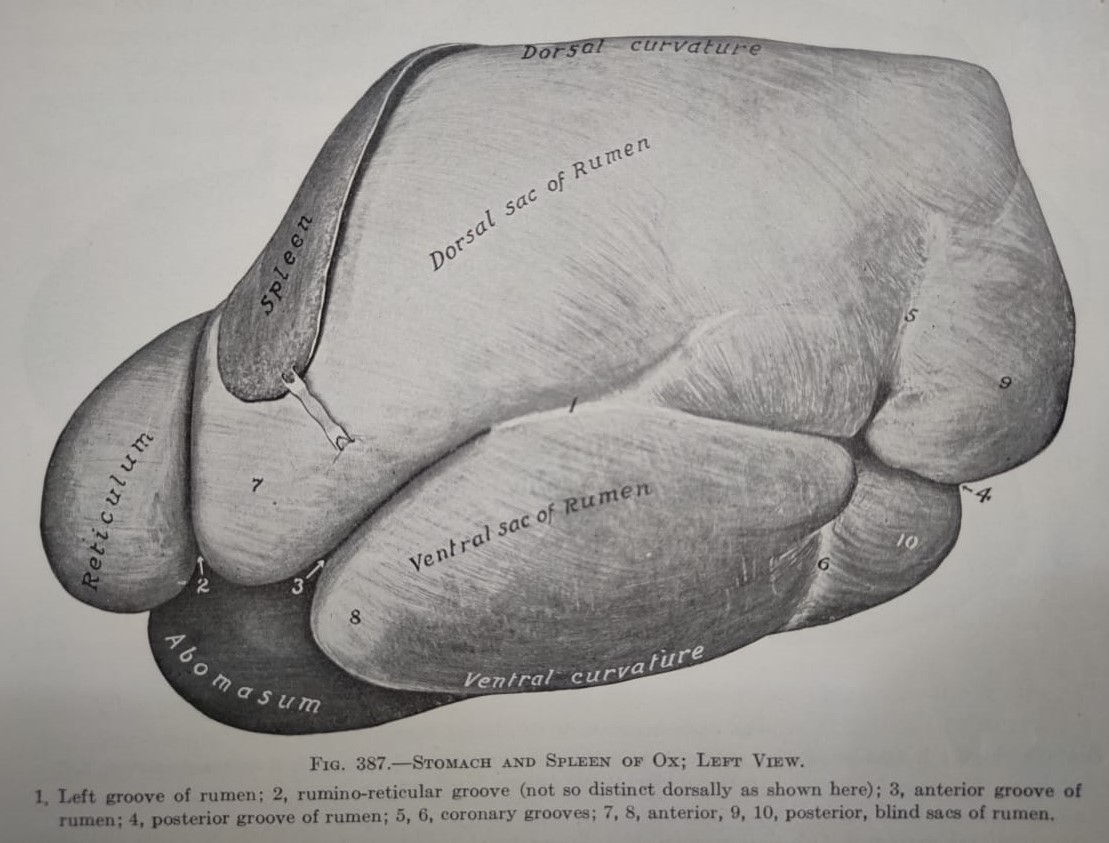
RUMEN
The rumen occupies most of the left half of the abdominal cavity and extends considerably to the right of the median plane ventrally and caudally. Its long axis reaches from a point opposite the ventral part of the 7th or 8th intercostal space almost to the pelvic inlet.
It has two surfaces, two curvatures
and two extremities.
Surface:
•
Parietal surface (left) – is convex and related to the diaphragm, the
left wall of abdomen and spleen.
•
Visceral surface (right) – is
somewhat irregular and is related chiefly to the omasum and abomasum, the
intestine, the liver, the pancreas, left kidney, left adrenal gland, the aorta
and the caudal venecava.
Curvatures:
•
Dorsal curvature – follows the curve formed by the
crura of the diaphragm and the sub-lumbar muscles; it is firmly attached to
these on the left by peritoneum and connective tissue as far caudad as the 4th
lumbar vertebra
•
Ventral curvature – lies on the floor of the abdomen,
the superficial wall of the omental bursa intervening.
Omental bursa–
- The omental bursa communicates with the peritoneal cavity by a relatively narrow passage termed the epiploic foramen(also known as foramen of winslow). This opening is situated on the visceral surface of the liver dorsal to the portal fissure. It's dorsal wall is formed by the caudate process and the caudal venacava. It's ventral wall consists of the pancreas, the hepato-duodenal ligament and the portal vein.
- This space is closed on the left by the stomach and the gastro-phrenic ligament, ventrally and on the right by lesser omentum, and dorsally by the gastro-pancreatic fold, which is attached to the dorsal border of the liver and to the caudal venacava.
- Thus the vestibule is closed except; (a) on the right, where it communicates with the peritoneal cavity by the epiploic foramen, and (b) caudally where it communicates with the caudal recess of the omental bursa.
Extremities:
•
Cranial extremities
–
is divided ventrally by a transverse cranial groove into two
sacs. The cranial sac is continuous caudally with dorsal sac of rumen and
cranially with the reticulum. It curves ventrally over the round cranial end of
ventral
sac. The external line of demarcation between the cranial sac and the
reticulum is the rumino-reticular groove. It is deep ventrally and is distinct
on part of the parietal surface, but dorsally no natural separation exists, the
rumen and reticulum together forming a dome like vestibule(rounded roof
like) on which the esophagus terminates.
•
Caudal extremities – extends nearly to the pubis and
is related to the intestine and bladder. It is divided into caudo-dorsal
and caudo-ventral
blind sacs by a deep transverse caudal groove which
connects the longitudinal grooves. The blind sacs are marked off by the dorsal
and ventral coronary grooves on each sides of the rumen.
The surfaces
are marked by the right and left longitudinal groove which
divides the rumen into dorsal and ventral sacs externally.
- On the right side, there are two grooves. The ventral one is the right longitudinal groove, extending from the cranial to the caudal groove, the dorsal one is the right accessory groove which is a curve, convex dorsally, and joins the right longitudinal groove at both ends, enclosing an elliptical area, the insula ruminis.
- The left longitudinal groove begins at the cranial groove, inclines at first dorsally and then ventrally, and joins the caudal groove. Near the middle it gives off a dorsal branch, the left accessory groove which extends caudo-dorsally and fades out.
The reticulum is the most cranial and smallest of the four
compartments. It is located between the 6th and 7th or 8th
ribs. The greater part of it lies on the left of the median plane. It is
somewhat piriform(pear shaped), but is compressed cranio-caudally.
It has two surfaces and two curvatures;
Surfaces:
• Diaphragmatic surface
• Visceral surface
The diaphragmatic surface- is convex and lies against the diaphragm.
The visceral surface- is
flattened more or less by the presence of atrium ruminis (cranial sac of
rumen).
Curvatures:
- The greater curvature
- The lesser curvature
• The greater curvature- faces to the left and ventrally;
it lies against the diaphragm opposite to the 6th and 7th
ribs.
• The lesser curvature- faces to the right and dorsally and is connected with the abomasum.
OMASUM
It is located opposite the 7th to 11th
ribs and lies chiefly to the right of the median plane. It is
ellipsoidal(oval/spindle) in form and somewhat compressed between it's parietal
and visceral surfaces.
It has two surfaces, a curvature and a base;
Surfaces:
- The parietal(right) surface
- The visceral (left) surface
The parietal(right) surface- faces obliquely to the right and cranially and is related chiefly to the diaphragm and liver.
The visceral (left) surface- faces in the opposite direction and in is contact with the rumen, reticulum and abomasum.
Curvature:
The
dorsal curvature- faces dorsally, caudally and to the right.
Base:
The base is very short and faces cranially, ventrally and to the left. It is connected to its upper part with the reticulum by a very short narrow neck of the omasum.
ABOMASUM
The abomasum
is an elongated sac which lies chiefly on the abdominal floor. The cranial
blind end, the fundus, is in the xiphoid region in relation to the reticulum,
to which it is in part attached, the cranial sac, and the ventral sac of rumen.
The body
extends caudally between the ventral sac of the rumen and the abomasum, lying
more on the left than on the right of the median plane.
The pyloric part turns to the right caudal to
the omasum, inclines dorsally and joins the duodenum at the pylorus, which is
usually at or near the ventral end of the 9th or 10th
intercostal space.
It has two surfaces
and two curvatures;
Surfaces:
•
Parietal surface
•
Visceral surface
•
The parietal surface- is in
contact mainly with the abdominal floor, while
•
The visceral surface- is for the most part related to
the rumen and the omasum.
Curvature:
•
The greater curvature
• The lesser curvature
The greater curvature- gives attachment to the superficial wall of greater omentum.
The lesser curvature- gives attachment to the lesser omentum, which passes over the parietal surface of the omasum to the liver. The abomasum and the omasum are also directly attached to each other by connective tissue. The abomasum in the sheep and goat is relatively larger and longer than in the ox.
Facebook Veterinary group
Dear DR. Anjani,
ReplyDeleteThank you very much for the good initiate. I appreciate your dedicated service.
All the best.
Post a Comment