Written By Anjani Mishra
 |
What is Ovulation
The defination of What is Ovulation :- The process of rupture of mature follicle(secondary oocyte) and discharge of ovum from the ovary is known as ovulation.
Mechanism of ovulation
- After formation of graafian follicle, it is pressed to one side of the surface of ovary by the pressure of liquor folliculi, where the epithelial surface of the ovary begins to bulge locally forming a projection known as stigma.
- Due to pressure of the ovum, the epithelial cells of this portion get less nutrition because of less blood supply. As a result, there is local weakening and degeneration of the ovarian surface.
- On the other hand increased intra-follicular pressure and muscular contraction in the ovarian wall, the ovum is released.
- Thus the oocyte together with surrounding granulosa cells from the region of cumulus oophorus breaks free and floats out of the ovary.
- At this moment, the oocyte with its cumulus oophorus cells is discharged from the ovary called ovulation.
Corpus
luteum- Corpus means
body, and
Luteum means
yellow means yellow body.
- After ovulation, the rupture follicle does not degenerate at once, but is transformed into temporary glandular structure called corpus luteum.
- During this process, the granulosa cells remaining in the wall of rupture follicle together with theca interna cells are vascularized by surrounding vessels and become polyhedral.
Fig: Ovary of bitch
(Showing several interstitial glands between two corpora lutea within the stroma)
1. Corpus luteum, 3. Granulosa lutein cell, 5. Interstitial gland
Formation of corpus luteum
After ovulation, several changes takes place during the formation of corpus luteum. These are as follows;
- The follicular cavity closes with serous fluid and blood.
- Membrana granulosa cells increase in size but they do not multiply in number.
- The theca interna cells increase in size as well as multiply in number.
- The theca externa cells merge with the ovarian stroma.
After these
changes membrana granulosa cells are converted into granulosa
lutein cells and the theca interna cells are converted
into theca
lutein cells.
- These two types of cells can be differentiated on the basis of their size, position and staining characters.
- The granulosa lutein cells are centrally in position and they are larger than theca lutein cells.
- The theca lutein cells are peripheral in position, smaller in size but their nuclei are stained darker.
- Cytoplasm of both lutein cells contain lipid droplets and pigments, so the cytoplasm of the cells appear vacuolated.
- Granulosa lutein cells secretes mainly progesterone and small amount of estrogen, where as theca interna cells secrete estrogen only.
- If the discharged ovum is not fertilized, then the corpus luteum reaches its greatest development about 1 week after ovulation and then begins to degenerate.
- The cells of such corpus luteum gradually decrease in size, show increase vacuolization and are finally reabsorbed as corpus albicans.
- If the discharged ovum is fertilized, the corpus luteum increases in size for a long time, which persist until the later months of pregnancy depends upon the development of placenta.
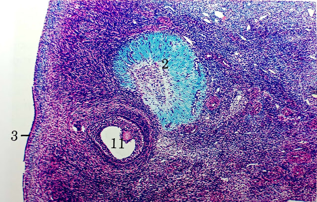
Fig: Ovary of cow, Corpus Albicans
(Showing scar tissue of corpus albicans, stained bright blue-green)
2.Corpus albicans, 3.Germinal epithelium, 11.Young tertiary follicle
Function
of corpus luteum
- It stimulates the endometrium to prepare the uterus for reception and nourishment of a fertilized ovum.
- It decreases the contraction of uterine muscles and prevents the expulsion of the developing embryo from the uterus.
- It prevent further ovulation by depressing the secretion of FSH from anterior pituitary.
What is Fertilization
The Defination of What is Fertilization :- is the process of fusion of two mature germ cells, an ovum and spermatozoan, to form a mono-nucleated single cell, the zygote.
- The fusion takes place usually in the ampullary region of the uterine tube. This is the widest part of the tube and is located close to the ovary.
- Spermatozoa can stay alive in the female reproductive tract for about 24 hrs., while secondary oocyte is thought to die within 12 to 24 hours after ovulation if not fertilized.
- Spermatozoa pass rapidly from the vagina into the uterus and subsequently into the uterine tubes.
- This ascent is caused by contraction of the musculature of the uterus and the uterine tube.
- It must be kept in mind that the spermatozoa upon arrival in the female genital tract are not capable of fertilizing the oocyte. They must undergo;
a) Capacitation,
&
b) Acrosomal
reaction
a) Capacitation
- Capacitation is a period of conditioning for spermatozoa in the female reproductive tract which, in the human, lasts approximately for 7hrs.
- During this time, a glycoprotein coat and seminal plasma proteins are removed from the plasma membrane that overlies the acrosomal region of the spermatozoa.
- Completion of capacitation permits the acrosome reaction to occur, where the only capacitated sperm can pass through the corona cells and undergo acrosome reaction.
b) Acrosome reaction
- Acrosome reaction occurs immediately after capacitation, where the plasma membrane and the outer acrosomal membrane fuse together, permitting the release of the acrosomal contents needed to penetrate the corona radiata and zona pellucida.
During acrosome
reaction the following substances are released;
i)
Hyaluronidase- needed to penetrate the corona radiata barrier,
ii)
Trypsin
like substance- needed for digestion of the zona pellucida, &
iii) Zona-lysin- needed to help the spermatozoa to cross
the zona pellucida.
The phases of
fertilization include;
1. Phase 1:
Penetration of corona radiata,
2. Phase 2:
Penetration of zona pellucida, &
3. Phase 3: Fusion
of oocyte and sperm cell membrane.
1
Phase 1
- Out of 200-300 million of spermatozoa deposited in the female genital tract, only 300-500 reach the site of fertilization where only one of those is needed for fertilization with one ovum.
2
Phase 2
- The zona pellucida is a glycoprotein shell surrounding the egg that facilitate sperm binding and induces the acrosomal reaction.
- Release of acrosomal enzyme allow sperm to penetrate the zona pellucida, thereby come in contact with the plasma membrane of the oocyte.
- Permeability of the zona pellucida changes when the head of the sperm comes in contact with oocyte surface.
- The contact results in release of lysosomal enzyme from corticol granules lining the plasma membrane of the oocyte, that cause an alteration in the properties of the zona pellucida (the zona reaction) to prevent further sperm penetration.
3
Phase 3
- As soon as the spermatozoa comes in contact with the oocyte cell membrane, the two plasma membrane fuse, since the plasma membrane covering the acrosomal head cap has disappeared during the acrosomal reaction.
- Actual fusion is accomplished between the oocyte membrane and the membrane that covers the posterior region of the sperm head.
- As soon as the spermatozoan has entered the oocyte, the egg responds in three different ways;
i) Cortical and zona reactions
- As a result of the release of cortical oocyte granules, the oocyte membrane becomes impenetrable to other spermatozoa as the zona pellucida alters its structure and composition.
ii) Resumption of the 2nd
meiotic division
- The oocyte finishes its 2nd meiotic division immediately after entry of the spermatozoan (from female pronucleus)
iii) Metabolic activation of the egg
- The activation factor is probably carried by the spermatozoan.
- The spermatozoan, mean while, moves forward until it lies in close proximity to the female pronucleus.
- It's nucleus becomes swollen and forms the male pronucleus.
The main results of fertilization are;
- Restoration of the diploid number of chromosomes, half from father and half from the mother.
- Determination of the sex of the new individual. An 'X' carrying sperm will produce a female (xx) embryo, and 'Y' produces a male (xy) embryo.
- Initiation of cleavage- without fertilization the oocyte usually degenerate after 24 hrs of ovulation.
If you have any questions you can ask me on:-
mishravetanatomy@gmail.com
Facebook Veterinary group link - https://www.facebook.com/groups/1287264324797711/
Twitter - @MishraVet
Facebook - Anjani Mishra
Website: mishravetanatomy.blogspot.com


Great tips regrading ovulation. You provided the best information which helps us a lot. Thanks for sharing the wonderful information.
ReplyDeletePost a Comment