What is Cleavage & What is Implantation ?
 | |
| Know about Cleavage and Implantation |
In
this post you are going to know about what is cleavage and implantation.
But
to know about cleavage and implantation, you first must know about ovulation
and fertilization.
So what is ovulation ?
Ovulation
is the process of rupture of mature follicle and discharge of ovum from the
ovary is known as ovulation.
Now,
you know about what is ovulation, So you must also know about what is
fertilization.
What is fertilization ?
Ferlization
is the process of fusion of two mature germ cells, an ovum and spermatozoan to
form a mono-nucleated single cell, the zygote.
For
more information about ovulation and fertilization, You can head to my last
post or click here :- Ovulation and Fertilization
Now,
Lets head to What is cleavage ?
What is Cleavage ?
Now lets start to know about cleavage
To
know about cleavage we need to know about it's Definition first.
Definition:
The series
of cell division that takes place immediately after fertilization
is called cleavage.
These daughter cells, which becomes smaller with each cleavage division, are known as blastomeres.
These daughter cells, which becomes smaller with each cleavage division, are known as blastomeres.
Once
the zygote has reached the two-cell stage, it undergoes a
series of mitotic cell divisions, resulting in a rapid increase
in the number of cells.
Do you know, What is zygote ?
The
mono-nucleated single cell fertilized ovum is known as zygote.
What is Morula (mulberry)
After
3 to 4 divisions the embryo, i.e., the blastomere cells increase
in number at certain stage, [approximately 3 days after fertilization] where
the cells are 16 in number and looks like a mulberry fruit is
called morula stage.
What does morula consists of ?
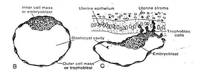
The morula cousists of a group of centrally located cells, the inner cell mass which gives rise to the development of embryo proper while surrounding cells of morula compose the outer cell mass, that forms the traphoblast which later contributes to the placenta. At this stage the embryo is about to enter the uterus.
Do you know how blastocyst is formed ?
Formation of blastocyst
When
morula enters the uterine cavity, fluid begins to penetrate through the zona
pellucida into the inter-cellular spaces of the inner cell mass.
Gradually,
the intercellular spaces become confluent, and finally, a single cavity, blastocele
is formed.
At
this stage, the embryo is known as blastocyst/blastula stage.
Cells
of inner cell mass, now referred to as the embryoblast, are
located at one pole, while the outer cell mass or trophoblast
becomes flattened and form the epithelial wall of the blastocyst.
What is Embryoblast ?
The inner cell mass that gives rise to the development of future axis of embryo is called embryoblast.
What is Embryoblast ?
The inner cell mass that gives rise to the development of future axis of embryo is called embryoblast.
The
zona pellucida now disappeared, allowing implantation to begin.
Now, You already knew about what is cleavage.
So, Let's know about what is implantation and it's process.
Now, You already knew about what is cleavage.
So, Let's know about what is implantation and it's process.
What is Implantation?
Fig: Implantation
Difinition : Implantation is the
process of attachment of blastocyst with or within the endometrium
of uterus.
Zona
pellucida
disappears at the end of 5th day of fertilization.
Implantation or embedding
of the blastocyst takes place on the 6th or 7th day after
fertilization.
Implantation is initiated
by the traphoblast cells, located over the embryoblast pole.
What is trophoblast ?
The outer cell mass of morula that contribute to form future placenta is called trophoblast.
The
trophoblasts are capable of secreting digestive/proteolytic
enzyme that dissolves the zona pellucida and cause penetration
and subsequent erosion of epithelial cells of the uterine mucosa.
The
uterine mucosa, however, promotes the proteolytic action of the blastocyst,
so that the cells of embryo can receive nutrition and oxygen from
uterine milk secreted by the uterine glands.
The
sticky nature of the milk help the embryo to
settle in the uterine pits and the process of implantation
begins for further development.
In
this way by the end of 1st week of development, the zygote has
passed through the morula and blastula stages and
has begun implantation in the uterine mucosa.
Formation
of germ layer
Bilaminar
germ disc:
At the 8th
day of development, the blastocyst is partially embedded in the endometrial
stroma. In the area over the embryoblast, trophoblast has differentiated into
two layers;
- An inner layer of mononucleated cells, the cytotrophoblast, and
- An outer layer of multinucleated zone or cells without distinct cell boundries, the syncytotrophoblast. These cells invade the endometrium of uterus and supplies nutrition to the embryo.
Cells of
inner cell mass or embryoblast also differentiate into two layers:
- A layer of small cuboidal cells adjacent to the blastocyst cavity is known as the hypoblast layer, and
- A layer of high columnar cells adjacent to the amniotic cavity is known as the epiblast layer.
Cells of each
germ layer form a flat disc and together are known as "bilaminar germ
disc".
Between
epiblast and hypoblast, there is an extra-cellular basement membrane that
separate the bilaminar germ disc.
At the same time,
a small cavity appears above the epiblast. This cavity enlarges to become the
amniotic cavity.
Those
epiblast cells adjacent to the cytotrophoblast cells are called amnioblast, and
together with the rest of the epiblast, line the amniotic cavity.
Fig: Cytotrophoblast and syncytotrophoblast cell
On
the other hand, the hypoblast encircle the unilaminar
blastocele forming bilaminar blstocele that gives rise to
the primary yolk sac.
Now
the bilaminar germ disc is formed between the primary amniotic
cavity above and the primary yolk sac below.
Trilaminar
germ disc
(3rd
week of development)
The
most characteristics events occurring during the 3rd week is the Gastrulation,
the process that establishes all three germ layers in the embryo, and these are
as follows;
A.
Ectoderm,
B.
Mesoderm, &
C.
Endoderm.
Appearance
of germ layers
At
first stage, the germ disc is circular, then it becomes
elongated, indicating the future axis of the embryo.
One
end becomes broad (cephalic end) and the other end becomes narrow
(caudal end).
Three functional
zones are differentiated in the ectodermal layer of the germ disc such as;
A. Surface ectoderm
B. Neural plate
C. Pluripotent cellular zone
A. Surface
ectoderm
The
cells of cephalic end and the peripheral margin constitute the surface
ectoderm, that gives rise to the epidermis of skin.
B. Neural plate
The
cells on the depressed area at the central part of the surface ectoderm form an
elongated plate, which gives rise to the future nervous system.
C. Pluripotent
cellular zone
A
group of fast proliferating cells are accommodated at the caudal part
of the germ disc.
They
differentiated very rapidly and form a linear opacity in the
midline, called primitive streak.
Gastrulation begins with
the formation of primitive streak on the surface of the
epiblast.
Initially
the cells of epiblast, i.e. pluripotent cells migrate towards the
primitive streak.
On
arrival in this region of the primitive streak, they become flask
shape, detach from the epiblast, and slip beneath it.
This
inward movement is known as invagination and this invagination
procedure between epiblast and hypoblast is known
as gastrulation.
Once
the cells have invaginated, some of them displace the hypoblast,
creating the embryonic endoderm, while some of them come to lie
between the epiblast and newly created endoderm to
form mesoderm.
Cells
remaining in the epiblast then form ectoderm. Thus
the epiblast, through the process of gastrulation,
is the source of all the germ layers and cells in these layers will give rise
to all of the tissues and organs in the embryo.
Therefore
all the three components of the tri-laminar germ disc, i.e. ectoderm,
mesoderm and endoderm are derived from pluripotent
epiblast by forming primitive streak.
Conclusion:
From
the above statement, it is concluded that immediately after fertilization,
the zygote undergoes a series of mitotic cell division results in
the formation of morula.
The
morula consists of centrally located inner cell mass that
gives rise to the embryo while the outer cell mass
contribute to the formation of placenta.
The
morula then enters the uterine cavity where fluid penetrate in the
intercellular space forming a single cavity called blastocele and
this stage is called blastocyst or blastula.
The
blastocyst gradually loses the zona pellucida allowing implantation
to begin which is initiated by trophoblast cells.
The
trophoblast cells by secreting proteolytic enzymes dissolve zona
pellucida and epithelium of uterine mucosa for attachment to establish
nutritional supply to the embryo.
After
implantation bilaminar germ disc is formed, initiated by inner
cell mass/embryoblast which is further differentiated into epiblast
and hypoblast.
The
epiblast cells give rise to the future ectoderm as
well as amniotic cavity above where as hypoblast
cells give rise to the future endoderm as well as the yolk
sac below.
With
the formation of bilaminar germ disc, the process of gastrulation
begins from ectoderm that gives rise to the
trilaminar germ disc.
The
ectoderm is further differentiated into three functional
zones as surface ectoderm, neural plate and pleuripotent
cellular zone.
The
pleuripotent cellular zone gives rise to the primitive streak and
with the formation of primitive streak, the process of gastrulation
begins that gives rise to the mesoderm.
In
this way, all the three components of the trilaminar germ
disc, i.e. ectoderm,
mesoderm,
and endoderm are derived from pleuripotent epiblast by forming primitive
streak.
Upon reading this post, I hope that you got to know about cleavage, Implantation as well as the development of bilaminar and trilaminar germ disc in animal.
Thank you.
Upon reading this post, I hope that you got to know about cleavage, Implantation as well as the development of bilaminar and trilaminar germ disc in animal.
Thank you.
If you have any questions you can ask me on :
mishravetanatomy@gmail.com
Facebook Veterinary group link - https://www.facebook.com/groups/1287264324797711/
Twitter - @MishraVet
Facebook - Anjani Mishra
Website: mishravetanatomy.blogspot.com


Post a Comment