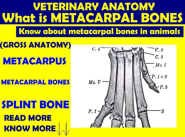
METACARPAL BONES
Written By Anjani Mishra

Metacarpal bones are the second section of manus and have
a large
metacarpal bone and an external small metacarpal bone on the lateral aspect of the proximal extrimity. The large metacarpal bone (Mc. 3+4) results from the fusion of the third and fourth bones of the foetus, and bears evidences of it double origin even in the adult stage. The shaft is shorter than in the horse, and is relatively wider and flatter.
Large Metacarpal bone
Shape:
It is long bone.
Location:
Placed in second section of manus between the carpus above and the digits
below.
Direction:
It is vertically placed.
Relation:
It is fused bone of the 3rd and 4th metacarpal bones.
Articulation: It articulates proximally with the distal row of carpal bones forming a carpal/knee joint and distally with the first phalanx forming a fetlock/metacarpo-phalangeal joint.
Articulation: It articulates proximally with the distal row of carpal bones forming a carpal/knee joint and distally with the first phalanx forming a fetlock/metacarpo-phalangeal joint.
Composition:
It has ONE shaft and TWO extremities.
Shaft:
It is semi-cylindrical and presents: 2 surfaces and 2 borders.
The anterior
surface is smooth and rounded from side to side, and present vertically
anterior vascular groove, which lodges two foramina, one proximal and other
distal. These foramina also communicate at posterior surface.
The posterior
surface is flat and is in contact with the suspensory ligament. There
is a shallow posterior vertical groove marking the union of the 3rd
and 4th metacarpal bones.
The borders are medial
and lateral,
which are smooth and rounded.
Extrimities:
Proximal
Extremity:
- It presents two slightly concave facets ( medial and lateral facets), which are separated by a median ridge in front and a deep notch behind.
- The medial facet is larger and articulates with the second and third fused carpal.
- Centrally, there is a synovial fossa.
- The lateral facet is smaller and articulates with 4th carpal.
- Just below this facet, on the lateral side, there is convex facet for articulation with the small metacarpal bone.
- There is metacarpal tuberosity, at the antero-medial aspect, to which the tendon of the extensor carpi radialis is attached.
- In front, there is a roughened elevation to which ulnaris lateralis is attached.
At the posterior aspect behind the articular margin, there is nodule for the insertion of flexor carpi radialis m/s
Distal Extremity:
- It is divided by the intercondyloid cleft into medial and lateral condyles, of which the medial lower in position than the lateral.
- Each condyle represents the distal extremity of one metacarpal bone.
- An antero-posterior ridge divides each condyle into two articular areas, the lateral of which higher than the medial.
- The condyle articulates with 1st phalanx and two sesamoid bones.
Small
Metacarpal Bones
- It is rod- like bone measuring about one and half to two inches in length and placed at the proximal part of the lateral border of the large metacarpal bone.
- The proximal extremity is rounded and rough while the distal extremity tapers to a point.
- The anterior surface presents proximally a small facet for articulation with the large metacarpal bone.
- Sometime, the bone is fused with the large metacarpal.
Comparison
with:
A)
Metacarpus of Horse:
In this region, there are 3 bones, one large metacarpal (3rd) and two small metacarpal (2nd and 4th). Of these, only the third one is well developed and carries a digit. The other two, the second and fourth are much reduced and are commonly called the small metacarpal or splint bone.
In this region, there are 3 bones, one large metacarpal (3rd) and two small metacarpal (2nd and 4th). Of these, only the third one is well developed and carries a digit. The other two, the second and fourth are much reduced and are commonly called the small metacarpal or splint bone.
large
metacarpal bone
- It is a long cylindrical and massive bone.
- The posterior surface is roughened on its dorsal two- third on either side for the attachment of the small metacarpal.
- The nutrient foramen is placed on the dorsal third of this surface.
- Anterior surface is smooth and convex.
- Anterior and posterior vascular grooves are absent.
- The proximal extremity present posterior, on either side, two facets, which articulate with the facets on the small metacarpal bones.
- The distal extremity is not divided and the whole articulate area of the lower extremity corresponds to one of the division of that of the ox.
- The distal extrimity is composed of two condyles, separated by a sagital ridge, of which the medial is slightly larger.
small
metacarpal bones:
- Are medial and lateral and extend to the distal-third of the large metacarpal bone.
- They represent the 2nd and 4th metacarpal bones.
- The lateral is slightly longer and slender where medial is slightly thicker/larger at its upper part.
- The proximal ends are larger and articulate with the large metacarpal while distal ends are pointed.
- The lateral small-metacarpal bone bears a single facet for articulation with the fourth carpal, while the medial bears two facets for articulation with the second and third carpal.
- The medial small metacarpal bone may present a small facet behind for the first carpal bone.
B)
Metacarpus of pig:
Four metacarpal bones are present.
Four metacarpal bones are present.
- The 1st is absent, the 3rd and 4th are larger and carry the chief digits, while the second and fifth are much smaller and bear the accessory digits.
- Their proximal end articulates with each other and with the carpus.
- Their distal end articulates with 1st phalanx and sesamoids.
Fig: Metacarpal of pig
Mc. 2(Metacarpal second), Mc.3(Metacarpal third), Mc. 4(Metacarpal fourth),
Mc. 5(Metacarpal fifth)
Mc. 5(Metacarpal fifth)
C) Metacarpus of Dog:
There
are five
metacarpal bones.
- The first is shortest.
- The second is slightly shorter than the 3rd and 4th, and a little longer than the 5th
- The 3rd and 4th are the longest.
- The 5th is the thickest of all the bones.
- The five bones articulate dorsally with each other by lateral facets, and with the corresponding carpal bones but diverge distally.
- Each metacarpal bone present a shaft and two extremities.
- It is nearly four sided in the third and fourth, three sided in the second and fifth, and is rounded in the first.
- The first has pulley-like lower articular surface while the other have convex surfaces.
Fig: Metacarpal bones of dog
Mc. I (Metacarpal first), Mc. V (Metacarpal fourth)
D)
Metacarpus of Goat:
- The metacarpal bones resemble those of the ox, except in size.
- The large metacarpal bone (3+4) is long slender.
- The lateral small metacarpal bone (Mc 5) is often absent or is presented by a ridge on the large metacarpal.
E)
Metacarpus of fowl:
- Have 3 bones (II, III, IV), which is fused to form a single mass.
- The 2nd metacarpal is a small projection while the 3rd and the 4th are fused at either extremities enclosing a narrow, elongated interosseous space between their shorts.
Fig: forelimb of chicken
11. Second metacarpal and Second digit. 12. Third metacarpal. 13. Third digit. 14. Fourth digit.
15. Fourth metacarpal. 16. ulna. 17. Radius. 18. Humerus.
15. Fourth metacarpal. 16. ulna. 17. Radius. 18. Humerus.
If you have any questions you can ask me on :
mishravetanatomy@gmail.com Facebook Veterinary group link - https://www.facebook.com/groups/1287264324797711/
Twitter - @MishraVet
Facebook - Anjani Mishra
Website: mishravetanatomy.blogspot.com
.......................................................................................................................................................................


Post a Comment