
Female Genital System of Animals


Ovary


- A central deeper portion is the medulla that consists of loose connective tissue, rich in elastic fibers and containing numerous large blood vessels, lymphatics, and nerves.
- The stroma contains scattered strands of smooth muscle fibers.
- The outer broad peripheral layer is the cortex, consists of a compact cellular stroma that contains the ovarian follicles.
- The stroma is composed of networks of collagen fibers and spindle shaped cells, arranged in irregular whorls. These stromal cells contribute to the growth of the theca folliculi.
- The follicles may be seen in all stages of development, and the appearance of the ovarian cortex depends upon the age of the individual and the stages of ovarian cycle.
- Before puberty only primary, or primitive follicles are seen. Sexual maturity is characterized by the presence of growing follicle and their end products (corpus luteum).
- After menopause, follicles disappear and the cortex eventually becomes a narrow region of fibrous connective tissue.
- An ovarian follicle consists of an oocyte surrounded by one or more layers of follicular cells, or granulosa cells.
- Ovarian follicles are embedded in the stroma of the cortex.
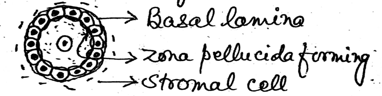
- A basal lamina underlies the follicular cell and marks the boundary between the follicle and the surrounding stroma.
- The follicles that are formed during fetal life are primordial follicles.
- It consists of a primary oocyte enveloped by a single layer of flattened follicular cells.
- The oocyte is a spherical cell about 25um in diameter. It has a large nucleus and nucleolus.


- As long as only a single layer of follicular cells encircles the oocyte, the follicle is called a "unilaminar primary follicle."
- At this stage, the glycoprotein rich zona pellucida begins to form between the oocyte and the follicular cells.
- A definite connective tissue capsule is absent around the follicle.
- The oocyte is a spherical cell about 30-40um in diameter.
- It has a large vesicular nucleus.
- Nucleolus is indistinct.
- Cytoplasm contains fine granules.
- Forming several layers of cells around the primary oocyte, the follicle is called “multilaminar primary follicle,” and follicular cells become to known as granulosa cells.
- During this stage, the zona pellucida thickens and the theca folliculi begins to form.
- The stroma immediately around the follicle differentiate to form the theca folliculi.
- A definite connective tissue capsule is present around the follicle.

- The multilaminar primary follicles continues to develop and increase in size, reaching upto 200um in diameter.
- Continued protiforation of the granulosa cells depends on follicle stimulating hormone (FSH).
- Irregular small spaces are found in the follicular mass, called antrum which is filled with fluid, called liquor folliculi.

- Continued proliferation of granulosa cells and liquor folliculi results in the formation of a mature (graafian) follicle.
- A relatively thick membrane is formed around the surface of the ovum known as zona pellucida.
- The ovum is pressed to one side of the follicle by the expansion of the antrum.
- The follicular epithelial cell immediately surrounding the ovum to form corona radiata.
- Granulosa cells surrounding oocyte remain intact and form the cumulus oophorus.
- The antrum is surrounded by a follicular epithelial cells, known as membrana granulosa.
- The theca externa gradually merges with the ovarian stroma.
- A connective tissue sheath is present around the follicular epithelium known as theca folliculi, composed of theca interna and theca externa.
- Theca folliculi separated from the membrana granulosa by a membrane, known as follicular basement membrane.


2. Sub-mucosa


2. Sub-mucosa- It consists of connective tissue together with large blood vessels, nerves and continuous with lamina propria as there is no muscularis mucosa.

- It is a thick layer of smooth muscle fibers, arranged both circularly and longitudinally.
- The muscularis(myometrium) layer is divided into thick inner circular and a thinner outer longitudinal layer invested by stratum vasculare, which contains numerous large blood vessels and nerves.
- The stratum vasculare is not very distinct in swine and man, where as in case of cow, located within the outer half of the circular muscle.
- I n some places particularly in the body and horn of uterus, the inner layer contains few oblique fibers and show reticular arrangement. During pregnancy, these muscle fibers increase in number and size under the influence of oestrogen.
- The stratum vasculare, and the layer of longitudinal muscle continues together with the broad ligament of uterus, which invests and suspends the uterus.
- The stratum vasculare corresponds to the sub-serosa. The longitudinal muscle layer should be interpreted as a muscularis serosae.


- The clitoris is rich in elastic fiber and consists of corpus cavernosum clitoridis, the glans, and the prepuce which continues with the vestibular mucosa.
- The corpus cavernosum clitoridis is homologous to the corpus cavernosum penis. It posses a heavy tunica albuginea with many vessels and nerves.
- The corpus cavernosum clitoridis of solipeds and women is well developed and equipped with abundant musculature.
- In ruminants there is less cavernosus tissue. In cat and sow varying quantities of fat cells are present in the trabeculae and cavernosus tissue. They are also found to a slight extend in solipeds and in larger quantities in ruminants.
- The end of the corpus cavernosum clitoridis is capped by the richly innervated glans. The glans clitoridis resembles the glans penis structurally.
- A true erectile body is present in the glans clitoridis only in the bitch and mare. Ruminants have only a fibroelastic cover on the distal end of the corpus.
- The preputium clitoridis, composed of the non-glandular cutaneous mucosa of the vestibule has parietal and visceral parts as in the male. It also contain lymph nodules. The space between these two layer of prepuce is the fossa clitoridis. The fossa is distinct in solipeds and bitch. In the ewe and cat there is only a slight indication of it, and it is absent in the cow and sow.
Estrus Cycle – The term estrus means
the time of strong mating tendency shown by a female. It is also properly
termed by breeders as heat.
A series of structural and physiological changes that take place in the
female reproductive organs from one estrus to another. The rhythmic changes
that take place in female from one estrus to another estrus is known as estrus
cycle.
The term estrus is derived from Greek Word “Oistrus” mean
Gadfly(mosquito), shows same behavior like estrus animal as cow.
The estrus cycle is
characterized by four distinct phases of changes as –
1. 1. Pro-estrus (18/19 - 21
days)
2. Estrus (0 -1 day)
3. Met-estrus (2 - 4
days)
4. Di-estrus (5 - 16,17,18 days)
The ovarian cycle
consists of two phases --
1. 1. Follicular phase
(proliferative stage)
Pro-estrus, estrus
2. 2. Luteal phase (secretory
stage)
Met-estrus, di-estrus
Facebook Veterinary group link - https://www.facebook.com/groups/1287264324797711/
Twitter - @MishraVet
Facebook - Anjani Mishra
I was childless for over 13 years after getting married, and the pressure from my mother-in-law nearly destroyed my marriage. Everyone believed I could never get pregnant, and my husband and I were constantly under stress. A colleague who had once been helped by Dr. Dawn during her own marriage struggles introduced me to him.
ReplyDeleteDr. Dawn performed a pregnancy ritual for me, and to my greatest surprise, within 3 weeks I conceived. My husband and his family began treating me like a queen—pampering me and showering me with love. Today, we are blessed with 3 beautiful children, all thanks to Dr. Dawn.
If you are facing relationship or marriage issues, or if you want to restore love, peace, and happiness in your family, I strongly recommend you contact Dr. Dawn—he will surely help you too.
His WhatsApp: +2349046229159
Email dawnacuna314@gmail.com
Post a Comment