Written By Anjani Mishra
Development of skeletal System
Axial skeleton
A.) Vertebral Column
Vertebral Column develops from paraxial
mesoderm. It divides into number of cubical blocks of tissue masses called somites or metameres. It is further divided into sclerotome and dermomyotome.
So, the sclerotome of the somites form the bones of vertebral column.
The cells of the sclerotome migrate
ventro-medially around the notochord.
Ø These migrated cells form a
segmented uniform but loose distribution at first.
Ø Soon at the middle of this segment, cells become compact in a transverse manner.
Ø This Condensed zone is called
perichordal disc, which subsequently form intervertebral disc.
Ø On either side of perichordal disc, the notochord disappears in less condensed areas.
Ø Now the less condensed area of two
adjoining segments face together to form the primordium of the body (centrum)
of a vertebra.
Ø The portions of the notochord which
did not disappear in the condensed zone expands to form nucleus pulposus.
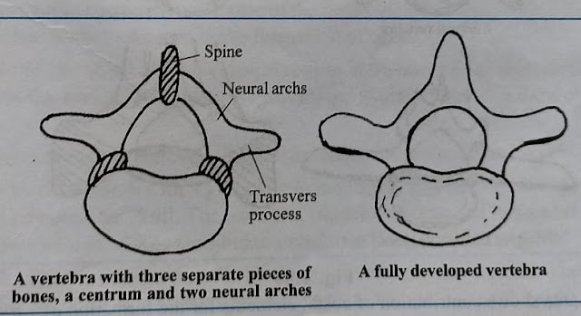
Ø The other cells of sclerotome
migrate dorsally and medially to form neural arches, spine and transverse
process.
(B) Rib
The ribs are formed from the condrification
of costal elements. These costal elements are developed in the neural arches
close to the centrum from separate centers.
In the thoracic regions, the costal
elements grow independently and form the costal arches.
First the arches becomes condrified
and subsequently ossified to form one primary ossification center. After birth
secondary ossification centers are developed for head and tubercle. The ventral
part remain cartilaginous.
C) Sternum
The ventral ends of cranial 7 or 8
costal arches fuse on each side to form cartilaginous sternal plate.
These plates unite at the midline in
cranio-caudal direction.
The fused cranial part form the manubrium, middle part forms the body and the caudal parts forms the xyphoid cartilage.
D) Skull
The occipital region at the base of skull is formed by the sclerotomes which are derived from occipital somites that appears in front of first cervical somite.
The temporal region is formed. by the
mesenchyme around the otic vesicles.
The first bronchial arch which gives
rise to maxillary and mandibular processes forms some facial bones of the skull.
The maxillary, palatine and part of
temporal bone develop from maxillary process, and the body of mandible from
mandibular arch.
The sphenoid bone develops from a
pair of polar cartilage appear by the side of pituitary gland. The ethmoid bone
is formed from a cartilaginous plate, the trabeculae cranii.
The nasal, lacrimal and vomer bones
develop from the membrane around nasal capsule.
The parietal and frontal bone develop
from mesenchyma surrounding the developing brain.
Development and Growth of bone
The primitive embryonal skeleton consists of cartilage and fibrous tissue, in which the bones develop. The process is termed ossification or osteogenesis, and is effected essentially by bone-producing cells, called osteoblasts. It is customary, therefore, to designate as membrane bones those which are developed in fibrous connective tissue, and as cartilage bones those which are preformed in cartilage. The principal membrane bones are those of the roof and sides of the cranium and most of the bones of the face. The cartilage bones comprise,therefore, most of the skeleton. Correspondingly we distinguish intramembranous and endochondral ossification. In intramembranous ossification the process begins at a definite center of ossification (Punctum ossificationis), where the osteoblasts surround themselves with a deposit of bone. The process extends from this center to the periphery of the future bone, thus producing a network of bony trabeculae. The trabeculae rapidly thicken and coalesce, forming a bony plate which is separated from the adjacent bones by persistent fibrous tissues.
The superficial part of the original tissue becomes periosteum,
and on the deep face of this successive layers of periosteal bone are formed by
osteoblasts until the bone attains its definitive thickness. Increase in
circumference takes place by ossification of the surrounding fibrous tissue,
which continues to grow until the bone has reached its definitive size. In
endochondral ossification the process is fundamentally the same, but not quite
so simple. Osteoblasts emigrate from the deep face of the perichondrium or
primitive periosteum into the cartilage and cause calcification of the matrix
or groundsubstance of the latter. Vessels extend into the calcifying area, the
cartilage cells shrink and disappear, forming primary marrow cavities which are
occupied by processes of the osteogenic tissue. There is thus formed a sort of
scaffolding of calcareous trabeculae on which the bone is constructed by the
osteoblasts. At the same time perichondral bone is formed by the osteoblasts of
the primitive periosteum. The calcified cartilage is broken down and absorbed through
the agency of large cells called osteoclasts, and is replaced by bone deposited
by the osteoblasts. The osteoclasts also cause absorption of the primitive
bone, producing the marrow cavities; thus in the case of the long bones the
primitive central spongy bone is largely absorbed to form the medullary cavity
of the shaft, and persists chiefly in the extremities. Destruction of the
central part and formation of subperiosteal bone continue until the shaft of
the bone has completed its growth.
mishravetanatomy@gmail.com
Facebook Veterinary group link - https://www.facebook.com/groups/1287264324797711/
Twitter - @MishraVet
Facebook - Anjani Mishra


Post a Comment