
Written by Anjani Mishra

Placenta
The placenta is a temporary physiological organ formed by the close association of the fetal membrane and the uterine lining, which permits the interchange of materials carried in the blood stream of the mother and the fetus.
Functions:
1. Nutrition-
CHO, protein, fat
2. Respiration-
O2 & CO2
3. Excretion-
Metabolic waste products
4. Barrier-
Prevent bac. & other antigen
5. Synthesis –Estrogen, progesteron
Placentation
The term placentation means the events which enable the embryo to get intimate relation with the uterus for its physiological causes or demand.
Classification of
placenta
A.
According
to the final distribution of the villi of the chorionic sac, the shape of the
placenta are classified into:-
1. Diffuse-
Uniform distribution of villi on placenta, i.e the chorionic villi are
distributed all over the chorion. Eg; sow, mare


2. Cotyledonary- If the villi concentrated in some area forming cotyledon, i.e. the villi are grouped in well spaced prominent cotyledon which are separated by smooth chorion. Eg; Cattle, Sheep, Goat


3. Zonary- Villi
concentrated at the centre of the placenta to form a belt like structure, i.e.
the villi are distributed and occupy a girdle like band about the middle of the
chorionic sac. Eg; Dog, Cat


4. Disoid- Villi
concentrated at one or two end of the placenta to form disc like structure i.e.
the villi are limited to one or two disc shaped area of the sac. Eg; Man,
Monkey

Chorionic frondosum
The
area of the chorion which attach with the maternal uterus is called chorionic
frondosum.
Chorionic leave
The
area of the chorion which is unattached and smooth part of the chorion is
called chorionic leave.
B.
On
the basis of degree of contact between the chorion and uterus and on the minute
microscopic relationship based on the number of tissue layers involved at the
zone of junction of the two components, i.e. in between the chorion and the
endomethium.
1.
Epithelio-chorial- The
epithelium of both fetal and maternal part remain intact found in diffuse type
of gross placenta. Eg; Mare, Sow

2.
Syndesmo-chorial-
All the components of the fetus remains intact but the epithelium of the
maternal part is destroyed found in cotyledonary type of gross placenta. Eg;
Cow, sheep, goat

3.
Endothelio-chorial-
All
the components of the fetus remains intact, but the epithelial tissue + C.T. of
the maternal part is destroyed found in zonary type of gross placenta. Eg; Dog,
cat, tigeress
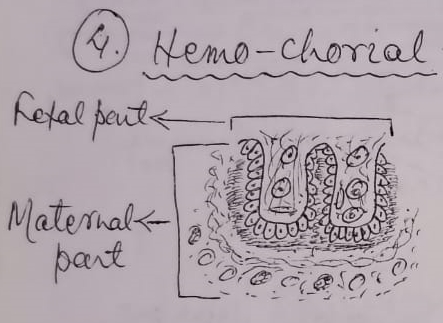
4.
Hemo-chorial-
All the components of the fetus remains intact, but the epithelial tissue +
C.T. + endothelium of the maternal part
is destroyed leaving the blood which comes in contact with the placenta. eg;
Man, Bat

5.
Hemo-endothelial-
The epithelial Tissue + C.T. of fetus is destroyed, where as epithelial Tissue
+ C.T. + endothelium of maternal part is destroyed leaving the blood which
comes in contact with the endothelium of the fetus. eg. Rabbit, guinea pig.


Fetal membrane
The
peripheral part of the germ layers contributing to the formation of certain
membranes, which serves for the purpose of protection, absorption of food,
respiration and excretion for the embryo. These membranes are not within the
body of the embryo and are discharged at the time of birth. These are also
called extra-embryonic membrane. The membranes are:-
1. Yolk
sac
2. Amnion
3. Chorion
4. Allantois

Fetal
membranes are formed by the following germ layers, as follows;
1. Ectoderm
2. Mesoderm-
Somatic and Splanchnic
3. Endoderm
The somatic membrane is very much close to the ectoderm called somatopleure and the splanchnic membrane is close to the endoderm called splanchnopleure.

1.
Yolk
Sac:
It
is also part of primitive gut which is not included within the body of the
embryo, when the embryo is folded off.
Function:
It supplies nutrients to the embryo by the formation of yolk material.

2.
Amnion:
It
arises as a layer of Somatopleure, which surrounds the developing embryo. In
the formation of amnion, the somatopleure thrown into folds, then gradually
come upward and after covering the embryo again comes downward and the limbs of
the both sides united ventrally.
The
inner limbs gradually forms a cavity called amniotic cavity containing amniotic
fluid and the outer limb becomes serosa which is gradually converted into
chorion.
Function:
i)
Protects the fetus from mechanical injury
ii)
Equalizes the pressure around the fetus
iii)
Prevent drying, adhesion and subsequent
malformation
3.
Chorion:
It
is the outermost layer of fetal membrane which encloses all other fetal
membrane including fetus. the outer limb of the somatopleure become serosa first
and gradually chorion.
Function:
It
forms the placenta and chorionic villi which is the source of nutrition,
respiration and excertion for the fetus.

4.
Allantois:
It
arises as a diverticulum from the hind gut of the embryo and gradually
increases in size and the yolk sac is gradually reduces. The wall of the
allantois is splanchnopleure. It is a slender endodermal tube which extends from
the caudal end of yolk-sac to the mesoderm of body stalk. When the wall of
allantois come in contact with the serosa, then the chorion is formed.
Function:
i)
It helps in maternal circulation
ii) It serves for the metabolic interchange between the fetus and mother.
If you have any questions you can ask me on :
mishravetanatomy@gmail.com
Facebook Veterinary
group link - https://www.facebook.com/groups/1287264324797711/
Twitter - @MishraVet
Facebook - Anjani Mishra
Website: mishravetanatomy.blogspot.com
Post a Comment