
Written by Anjani Mishra

Eye
The eye or organ of vision in the broader sense of the
term, comprises the eye-ball or globe of the eye, the optic nerve and certain
accessory organs associated there with. The accessory organs are the orbital
fasciae, muscles of eye-ball, the eyelids, conjunctiva and the lacrimal
apparatus.
The eye is special organ of the sense of sight. It is
situated in the orbital cavity. It is smaller in diameter than the orbital
cavity and the intervening space is occupied by the fatty tissue. The bony walls
of the orbit and the fatty tissue help to protect the eye from injury.
Structurally the two eyes are the same, some of their activities are coordinated, so that they function as a pair. They are supplied by the optic nerve.

Structure
There are three layers of tissue which constitute the
wall of the eye. They are;
1. The
outer fibrous layer: sclera and cornea
2. The
middle vascular layer: choroid, ciliary body & iris
3. The
inner nervous tissue layer: retina
The other structures includes;
4. The
lens
5. The
aqueous humor
6. The
vitreous body
The accessory structures include;
1. The
orbital fascia
2. The
muscles of eye-ball
3. The
eyelids
4. The
conjunctiva
5. The lacrimal apparatus

1. The
outer fibrous layer
The sclera and cornea
The
sclera: the sclera of the eye is composed of fibrous tissue
which consists of mainly collagenous fiber with some elastic fiber. It is
opaque and forms the outer layer of tissue of the posterior and lateral aspects
of the eye-ball and is continuous anteriorly with the transparent cornea. It is
a firm membrane and maintain the shape and form of the eye. It gives attachment
to the extrinsic muscles of the eye.
The
cornea: the cornea forms the anterior fifth of the fibrous
tunic. It is transparent, colourless and non-vascular except at its periphery
but posseses a well developed plexus of nerve. The light rays pass through the
cornea to reach the retina. It is convex anteriorly and involved in the
refraction or bending of light rays to focus them on the retina.
2. The
middle vascular layer:
The choroid, ciliary body & iris
The
choroid: the choroid is a thin membrane which lies between the
sclera and retina. It is very rich in blood vessels and is a deep chocolate
brown in colour. Posteriorly the choroid is perforated by the optic nerve and
anteriorly it is continuous with the ciliary body.
The
ciliary body: the ciliary body is a continuation of the
choroid anteriorly, consisting of non-striated muscle fibers (ciliary muscle)
and epithelial cells. It gives attachment to a fine ligament called suspensory
ligament, which at its other end is attached to the capsule of the lens. The
contraction and relaxation of the ciliary muscle changes the thickness of the
lens which bends the light rays entering the eye to focus them on the retina.
The epithelial cells secrete aqueous fluid into the
anterior segment of the eye, that is the space in front of the lens between
iris and cornea. It may also be associated with the secretion of the vitreous
body which occupies the space behind the lens.
The
iris: the iris is a muscular diaphragm of the eye, which
extends anteriorly from the ciliary body covering the anterior one-sixth of the
eye. It lies behind cornea and in front of the lens. It divides the anterior
segment of the eye into anterior and posterior chambers which contain aqueous
fluid. It is a circular body composed of pigment cells and two layers of muscle
fibers, one circular and the other radiating. In the center, there is an
aperture called pupil. The pupil varies in size depending upon the intensity of
light present. In the bright light the circular muscle fibers contract and
constrict the pupil. In dim light the radiating muscle fibers contract and
dilate the pupil. The iris is the coloured part of the eye and its colour
depends on the number of pigment cells present.
3. The
inner nervous tissue layer:
retina
The retina: the retina is the innermost layer layer of the wall of the eye. It is an external delicate membrane and specially adapted to be stimulated by light rays. It is composed of several layers of nerve cells and nerve fibers lying on a pigmented layer of epithelial cells which attached it to the choroid. It is described as the photosensitive part of the eye. The light sensitive cells in the retina are the rods and cones. Light rays cause chemicals changes in these cells and they emit nerve impulses which pass to the optic nerve.
Accessory
structures
The
orbital fascia: the orbital fascia surrounds the contents
of the orbit. It consists of three layers; the outer, middle and the deepest.
The outer layer arises at the periphery of the optic foramen and extends
forward to the eye lids; it sends septa between the eye muscles. This fascia is
just under the periorbital and is thin.
The middle/second layer of fascia originates in the
vicinity of the optic foramen and the orbital fissure. It also consists of two
layers; the outer and inner layer.
The deepest/third layer of fascia is called Tendon’s
capsule. This layer may be traced from the limbus area over the bulb and
closely covers the retractor bulbi muscle to the optic foramen.
The
muscles of eye ball: the eyes of domestic animals move within
their orbits, and this movement results from the action of the extraocular
muscles. All domestic animals, with the exception of the avian species, have
the same extraocular muscles. However, the sizes of the muscles, the exact
points of attachment and the exact innervation may show some slight difference.
There are seven extrinsic bulbi muscles; 4 rectus, 2
oblique and 1 retractor.
1. Rectus
dorsalis, ventralis, medialis and lateralis
2. Oblique
dorsalis and ventralis
3. Retractor
bulbi


Fig: Muscles of eye-ball (extrinsic)
The eye lids: these are two fibrous sheets, attached to the periphery of the orbital margin which cover the anterior portion of the eye-ball when it is closed. The slit between them is known as palpebral fissure. They protect the eye, prevent drying of the cornea by distributing lacrimal secretions and contain glands that assist in lubricating the cornea and specially the margin of the lids. The outer surface of the lids is covered with stiff hairs termed cilia or eye lashes. The eye lids are composed of skin, subcutaneous areolar tissue, fibers from orbicularis oculi muscle and conjunctiva.

The
conjunctiva: the membrane lining the eye lid and
covering the eye-ball is known as conjunctiva. It is a thin transparent
membrane which covers the front portion of sclera and cornea and is reflected
to the inner surface of palpebral at the palpebral fissure. The portion lining
the eye lid is called palpebral conjunctiva and the portion covering the eye-ball
is known as bulbar conjunctiva. The line of reflection is known as fornix
conjunctiva. When the eye lids are in apposition, the conjunctiva encloses a
capillary space between the lids and eye-ball and is known as conjunctival sac.
It protects the delicate cornea and the front of the eye.
The
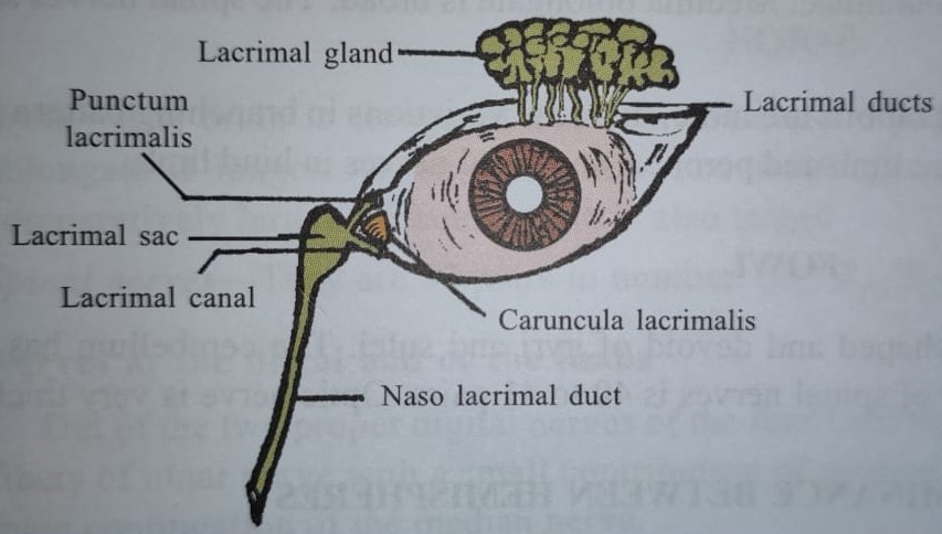
lacrimal apparatus: the lacrimal apparatus comprises
following structures;
Lacrimal and accessory gland
Lacrimal duct or excretory duct
Caruncula lacrimalis
Punctum lacrimalis
Lacrimal canals
Lacrimal sac
Naso-lacrimal duct
Lacrimal
gland: The gland is situated below supraorbital process and
inside of the upper lateral margin of the orbital cavity. The gland is
lobulated and tubulo-acinar in structure. The secretion is conveyed through
excretory duct (about 12 in number) to the superior conjunctival fornix. There is
a triangular space at the medial angle of the eye known as lacus lacrimalis.
Caruncula
lacrimalis: is a small rounded body situated in the
lacus and helps in the flow of tears in proper direction.
Puncta
lacrimalis: are minute orifices of the lacrimal canal
at the lacrimal papilla, situated on the margin of each lid and close to the
lacus lacrimalis.
Lacrimal
canals: are two in number; superior and inferior. They connect
lacus lacrimalis to the lacrimal sac.
Lacrimal
sac:
The sac is lodged in a fossa at the front end of the medial wall or the orbit.
Nasolacrimal
duct: starts from the sac, passes through the nasolacrimal
canal and opens on the external wall of the nostril.
Facebook Veterinary group
Best of luck dear friend.
ReplyDeleteProud of your efforts..
Post a Comment