
Written by Anjani Mishra

Iliac artery-External and internal iliac arteries
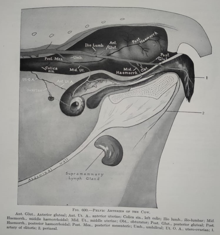
Internal
iliac arteries
Are large vessels that pass caudally and somewhat laterally
ventral to the wing of the sacrum towards the pelvic cavity.
Origin-
It arises or bifurcates under the 5th or 6th lumbar
vertebra.
Branches-
- Umbilical
artery
- Iliolumbar/iliomuscular
artery
- Cranial
gluteal artery
- Uro-genital
artery
- Caudal
gluteal artery
- Obturator
artery
- Internal pudendal artery
1. Umbilical
artery
Origin- arises
from the ventral surface of the internal iliac artery. It is very large vessels
in fetus.
Branches-
a. The
diferential artery- supplies ductus deferens in the male
b. The
uterine artery- supplies the uterus
c. The
ureteric artery- supplies the ureter
d. The cranial vesicular artery- supplies the cranial aspect of urinary bladder
2. Iliolumbar/iliomuscular
artery- psoas major and iliacus muscle
3. Cranial
gluteal artery- gluteus medius, profundus, gluteobiceps
4. Uro-genital
artery- (male and female)
Male
a. The
caudal vesicular artery- caudal aspect of urinary bladder
b. The
prostatic artery- prostate gland
c. The
urethral branch- major part of pelvic urethra
i.
Middle rectal artery- spincter ani
externus
ii. Dorsal perineal artery- skin of perineal region
Female (divides into cranial and
caudal branch)
a. Cranial
branch
i.
The uterine branch
·
The caudal vesicular artery- neck of
urinary bladder
·
The urethral branch- major part of pelvic
urethra
b. Caudal
branch
i.
Dorsal perineal artery- rectum and
clitoris
ii.
Caudal labial branch- vulva
iii. Caudal rectal artery- caudal segment of rectum
5. Caudal
gluteal artery- gluteobiceps, gemelli muscles
6. Obturator
artery- intra-pelvic part of obturator externus and adductor
7. Internal
pudendal artery (male and female)
Male
a. The
caudal rectal artery- wall of rectum
b. The
ventral perineal artery- perineal region
c. The
artery of the penis
i. The artery of the bulb- bulb of penis
ii. The deep artery of the penis- corpus
cavernosum penis
iii. The dorsal artery of the penis- dorsum of
the penis to the glans
Female
a. Ventral
perineal artery- cutaneous branch of perineal region, mammary gland
b. Artery
of the clitoris
i. Deep artery of the clitoris- crus of
clitoris
ii. Dorsal artery of the clitoris- clitoris

External
iliac arteries
Origin- Arises
from the abdominal aorta ventral to the body of the 6th vertebra,
but it may separate at the junction of the 4th and 5th
lumbar vertebra in ox.
Branches-
- Deep circumflex iliac artery
- Deep femoral artery
- Lateral circumflex femoral artery
- Caudal femoral artery
- Genicular artery
- Saphenous artery
- Popliteal artery

1. Deep circumflex iliac artery- abdominal
muscles
2. Deep
femoral artery
a. Pudendoepigastric
artery
i.
The caudal deep epigastric- obliquus
internus, rectus abdominis
ii.
The
external pudendal artery
Male-
scrotum, prepuce, penis, tunica vaginalis
Female- mammary gland
b. The medial circumflex femoral artery
3. Lateral
circumflex femoral artery
a. The
ascending branch- vastus lateralis, rectus femoris, iliacus, tensor fascia
latae
b. The descending branch- rectus femoris, vastus medialis, vastus intermedius
4. Caudal femoral artery- gluteobiceps, vastus lateralis, flexor digitorum superficialis
5.
Genicular artery- Sartorius,
semimembranosus, quadriceps femoris, vastus medialis, vastus intermedius
6.
Saphenous artery
a. The
medial planter artery- fascia and skin of tarsus
b. The
planter metatarsal artery- metatarsal region
c. Planter
common digital artery- digits region
7.
Popliteal artery
a. Cranial
tibial artery- tibialis cranialis
b.
Caudal tibial artery- flexor digitorum
profundus and superficialis, popliteus

- Continuation of the abdominal aorta in the sacrocaudal region. It is about 5 mm in diameter.
- It arises as an unpaired vessel from the dorsal aspect of the abdominal aorta between the two internal iliac arteries.
- It courses caudally along the pelvic surface of the sacrum and, beyond the first coccygeal vertebra continues as median coccygeal (caudal) artery.
- It releases paired segmental branches that pass through ventral sacral foramina to supply the meninges and spinal cord and emerge through the dorsal sacral foramina supplying the epaxial muscles of the sacrocaudal region.
- The last sacral branch of both sides may arise together by a common trunk. It passes dorso-caudally between the last sacrum and first caudal vertebrae and giving off the dorsal and ventral branch that supplies the muscles of this region.
- At about the level of first caudal vertebra the median sacral artery continues as the median caudal artery along the ventral surface of the entire length of the tail.
- It courses inside the vascular groove enclosed by the hemal processes, which sometimes fused, forming hemal arches.
- At regular intervals along its course, it releases paired segmental branches. They arise near the middle of each caudal vertebra and course dorsally and caudally, giving off the ventral and dorsal branches.
- These branches anastomose with the corresponding adjacent ones constituting the ventrolateral caudal and dorsolateral caudal arteries. They course along the ventral and dorsal aspect of the transverse processes of the caudal vertebrae.
- After giving off the ventral and dorsal branches, the caudal branches extend dorsally, supplying the dorsal muscles of the tail.
Facebook Veterinary group
Post a Comment