
Pelvic Girdle/Bony Pelvis
Written By Anjani Mishra
The bones of hind limb consists of four segments
1. Pelvic girdle/Bony pelvis:
Hip bone/Ossa Coxarum and sacrum
Hip bone consists of os coxae of both sides
Os-coxae consists of three bones on each side
Hip bone/Ossa Coxarum and sacrum
Hip bone consists of os coxae of both sides
Os-coxae consists of three bones on each side
Ilium, Ischium,
and Pubis
2. Thigh:
Femur
3. Leg/Crus
Tibia/Fibula
4. Pes/ Hindpaw
Tarsus(hock/ankle)
– Tarsal bones
Metatarsus –
Metatarsal bones
Digit/digits –
Phalanges and Sesamoid bones 1. PELVIC GIRDLE (BONY PELVIS)


The
PELVIC GIRDLE (BONY PELVIS)
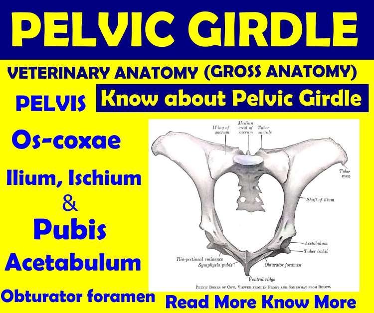
- The PELVIC GIRDLE consists of two similar bones, the os-coxae of both sides and the sacrum.
- Each os-coxae consists of three bones, namely; ilium, ischium, and pubis which are fused ventrally at symphysis pelvis/pelvic symphysis forming ossa-coxarum.
- Symphysis pelvis consists of ischial symphysis anteriorly and pubic symphysis posteriorly.
- Ossa-coxarum consists of two two ox-coxae of each side,which form a cartilaginous joint along the median line(pelvic symphysis).
PELVIS
The bony pelvis is composed of ossa-coxarum, the sacrum and the first few coccygeal/caudal vertebrae.
The bony pelvis(similar to basin) is bounded by pelvic bones and encloses a space called the pelvic cavity.
The cavity is simple ovoid and is free in communication with the abdominal cavity in front.
The floor and the roof of the pelvic cavity are not correspondingly placed. The floor or ventral wall is formed by the pubis and ischium bones and the roof or dorsal wall is formed by the sacrum and first few coccygeal vertebrae. The lateral walls are formed by the parts of ilium, sacro-sciatic ligament and acetabular part of ischia. .
Anterior part of the roof has no bony floor, hence it is called false pelvis.
The cranial opening of the pelvis is known as pelvic inlet or anterior aperture, which is formed dorsally by the sacrum base(sacral promontory), laterally by the shaft of the ilia(arcuate line/ilio-pectinal line) and ventrally by anterior border of the pubis(pecten ossis pubis).
The pelvic inlet has two principal diameter. The conjugate or sacro-pubic diameter, measured from the sacral promontary to the pecten ossis pubis. The transverse diameter is measured at the greatest width, i.e., just dorsal to the psoas tubercle.
The pelvic outlet or posterior aperture is smaller and incomplete at the sides which is bounded dorsally by the third or fourth coccygeal vertebra, and ventrally by the ischial arch and laterally by the broad sacro-tuberal ligament and semi-membranosus muscle, thus enclosing perinium.

OS COXAE/HIP BONE
- The os-coxae or hipbone is the largest of the flat bones.
- It consists of 3 bones primarily, the ilium, ischium, and pubis which meet together to form the acetabulum or cotyloid cavity, on each side which articulates with the head of femur.
- These bones are fused in the adult usually by 7 to 10 months, but it is convenient to describe them separately.
- It is a flat irregular bone, being directed obliquely downward and backward.
THE ILIUM:
- It is smaller than ischium and is irregularly triangular in shape.
- It is present at the cranio-lateral aspect of the pelvis.
- The bone is flat and expanded above, narrow in the middle and slightly expanded below.
- The narrow part is the shaft of the ilium and is short and flattened from side to side.
- The wide proximal part of the bone is called wing of the ilium.
Surfaces:
A) Gluteal Surface
B) Iliac/Anterior Surface
C) Pelvic/ Sacral surface
A)
Gluteal Surface:
- Gluteal surface faces upward and outward.
- It is concave at the proximal part and concavo-convex ventrally.
- Parallel to the lateral border, there is a prominent oblique ridge called gluteal line, which becomes continuous ventrally with superior ischiatic spine.
- Nutrient foramen is placed on the gluteal line close to the posterior border.
- Medial & deep gluteus muscle is attached to this surface.
B)
Iliac/ Anterior Surface:
- This surface faces forward, and is smooth and covered by iliacus muscle.
- Just above the acetabulum, there is deep depression for the attachment of the rectus femoris muscle.
C)
Pelvic/ Sacral Surface:
- This surface faces inwards, towards the pelvic cavity.
- Surface is wider at proximal and distal parts, but is narrow in the middle.
- The proximal wider part posses a triangular articular facet, which join with sacrum, forming sacro-iliac articulation. The margin of this facet is rough for attachment of sacro-iliac ligament.
Borders:
A) Lateral / Cotyloid Border
B) Dorsal Border
C) Medial Border
a)
Anterior / Public Border
b)
Posterior / Ischiatic Border
A)
Lateral / Cotyloid Border:
- It is concave, rough, separates the gluteal & the iliac surfaces and reaches the cotyloid cavity.
B)
Dorsal Border:
- It is thick, rough, concave and irregular, and forms the iliac crest on either end forming an angles.
C)
Medial Border:
- It is concave and at the beginning of the shaft, it is divided into anterior(public) and a posterior(ischiatic) border.
a)
Anterior/Public border/Ilio-pectinal line:
- It is rounded concave ridge.
- It begins in front and below the articular facet and joins the anterior border of the pubis.
- It separates iliac and pelvic surfaces and forms the lateral boundary of the pelvic brim.
- About middle of this line, there is psoas tubercle on which psoas minor muscle is inserted.
b) Posterior/Ischiatic Border:
- It is deeply concave and forms about its middle, the anterior boundary of the greater schiatic foramen through which nerves and anterior gluteal vessel passes.
- At its lower area, above the level of cotyloid cavity, this border is raised to form a part of superior ischiatic spine, which gives attachment to sacro-sciatic ligament.
- It separates the gluteal and the pelvic surfaces and is continued on the ischium.
Angles:
A) External angle / Tuber coxae
B) Internal angle / Tuber sacrale
C) Ventral angle
A)
External angle / Tuber coxae
- It is very large, prominent and is compounded of the three tuberosities.
B)
Internal angle / Tuber sacrale
- It is a little below the level of the sacral spines and lies opposite to the first sacral spine.
C)
Ventral angle
- It is the lower extremity of the bone.
- It meets the ischium and pubis at cotyloid cavity.
THE ISCHIUM:
- It is larger in size than ilium and is placed behind the ilium and the pubis.
- It is directed obliquely upward and backward.
- The transverse axis is pointing downward and inward; hence the pelvic floor is deeply concave.
It presents 2
surfaces, 4 borders & 4 angles.
Surfaces:
A) Pelvic Surface
B) Ventral Surface
A)
Pelvic Surface:
- It is smooth and concave and forms the posterior part of the pelvic floor.
- Just behind the anterior border, which forms the posterior margin of the obturator foramen, there is smooth, wide and less distinct groove for the attachment of the tendon of obturator internus muscle.
B)
Ventral Surface:
- It is nearly flat and presents a less developed, curved ridge about its middle, which extends either from the ventral surface of the tuber ischii or from the middle of this surface to terminate into a tubercle.
- Biceps femoris muscle originates on the ridge and the tubercle.
- The area lateral to the ridge, presents muscular imprints for the origin of gemelli muscles.
Borders:
A)
Anterior Border
B)
Posterior Border
C)
Medial Border
D)
Lateral Border
A)
Anterior Border:
- It is concave and forms the posterior margin of the obturator foramen.
B)
Posterior Border:
- It is thick and rough. It slopes inward and forward to meet the border of opposite side forming the ischial arch.
C)
Medial Border:
- It meets the opposite bone at symphysis ischii, which ventrally marks a roughened ridge terminating anteriorly into a tubercle.
D)
Lateral Border:
- It is thick smooth and concave, and forms the lesser sciatic notch over which passes the posterior gluteal vessels.
Angles:
A)
Antero-Internal
Angle
B)
Antero-External
Angle
C)
Postero-Internal
Angle
D)
Dostero-External
Angle
- The symphyseal branch of the ischium meets the pubis at this angle, and with it forms the internal boundary of the obturator foramen.
- The acetabular branch of the ischium meets the ilium and pubis at this angle in the cotyoid cavity.
- This angle meets the same angle of the opposite bone at the ischial arch.
- It is rough and thick.
- It forms trifid process (three sided mass) called tuber ischii, to which the sacro-sciatic ligament, biceps femoris, semitendinosus, semimembranosus and ischio-cavarnosus muscles are attached.
THE PUBIS:
- It is smallest bone of os-coxae and is placed between the ilium and the ischium.
- It is irregularly triangular and forms the anterior part of the pelvic floor.
It
presents 2 surfaces, 3 angles and 3 borders
Surface:
A)
Pelvic
Surface
B)
Ventral
Surface
A)
Pelvic Surface:
- It is smooth and rounded & large urinary bladder is placed on it.
B)
Ventral Surface:
- It is smooth and slightly convex.
Borders:
A)
Anterior Border
B)
Medial Border
C)
Posterior Border
A)
Anterior Border:
- It presents an oblique sub-pubic groove.
- Lateral of this border, there is ilio-pectinal eminence, to which the common pre-public tendon is attached that gives insertion to the abdominal muscles(e.g., obliq.ext. abdominis & rectus abdominis muscle).
B)
Medial Border:
- It joins the same border of opposite bone at the symphysis pubis.
C)
Posterior Border:
- It is concave and forms the anterior boundary of the obturator foramen, and is marked laterally by the obturator groove.
Angles:
A) Antero-Internal
Angle
B) Antero-External
Angle
C) Posterior
Angle
A)
Antero-Internal Angle:
- It is opposite to the same angle of opposite bone at the anterior end of the symphysis pubis.
B)
Antero-External Angle:
- It joins the ilium and ischium at the acetabulum.
C)
Posterior Angle:
- It meets antero-internal angle of the ischium, with which it forms the inner boundary of obturator foramen.
The Acetabulum:
- The acetabulum/cotyloid cavity is formed by the union of ilium, ischium and pubis.
- It articulates with the head of femur.
- It is directed outward and downward, and consists of articular and non-articular area.
- The articular part is divided into lateral and medial part.
- The non-articular part(accetabular fossa) is cut into its depth by a rough non-articular depression called acetabular notch.
- Acetabulum is surrounded by a rim to which a ring of fibro-cartilage is attached.
- It presents three notches; namely: postero-internal (acetabular) notch, antero-internal notch & external notch.
- The postero-internal notch is deeper and leads into acetabular fossa.
- The antero-internal and external notches are small and shallow and don’t reach to the acetabular fossa.
The Obturator Foramen:
- The obturator foramen is a large oval opening found on the floor of the pelvis and is formed by the ischium and the pubis.
- It is the largest foramen of the body.
- The margin is thin and sharp, except at its external parts.
- Its long axis is directed outwards and forwards.
- In the fresh state, the foramen is closed by fibrous membrane, ligaments and muscle ( obturator externus & obturator internus muscles), leaving a narrow space for passage of vessels and nerves.
DIFFERENCES IN MALE AND FEMALE HIP BONES:
Marked differences exist in size and form of the
pelvis of the two sexes.
- Both transverse diameter ( distance between two psoas tubercles) and conjugate diameter(length between body of sacrum and cranial end of the public symphysis) are greater in female.
- Inclination of the pelvis towards front, is greater in female.
- Pelvic outlet and ischial arch are wider in female.
- The angle made by symphysis ishcii, are greater in female and makes the pelvic cavity roomier in female.
- The obturator foramina are larger in the female.
Comparison
with:
A)
Pelvic Bone of Horse:


- The ischium is placed more obliquely.
- The gluteal line is faint.
- The nutrient foramen is placed on or near the posterior part of lateral border.
- The two-ischii meet at a greater angle, rendering the pelvic floor like bowl/basin.
- The inferior ischiatic spine, which is present only in horse, runs from the under surface of the tuber ischia inward and forward.
- The acetabulum and acetabular notch is wider than ox. The secondary acetabular notch is absent.
- The superior ischiatic spine is not so sharp.
- The tuber ischia is prominent but doesn’t present the trifid process as in ox.
- The subpubic groove is wider, more extensive and well marked.
- Psoas tubercle is less developed.
- The obturator foramen is small.
B)
Pelvic Bone of Dog:


- The ilium is nearly vertical and iliac shaft is compressed from side to side.
- The gluteal surface of ilium is more concave and is directed directly outwards.
- The pelvic surface is nearly flat.
- The dorsal border is convex, thick and rough.
- The pubic border is better marked and is continuous.
- The external angle is undivided.
- The superior ischiatic spine is low but thick.
- The greater ischiatic notch is shallow.
- The lesser ischiatic notch is absent.
- The ischium has a twisted appearance and ischial arch is very wide.
- The sub-pubic groove is absent.
- The acetabulum and acetabular fossa are deep.
- The acetabular foramen is triangular in outline with the angles rounded off.
C)
Pelvic Bone of Pig:


- Ilium is less extended laterally and dorsal border is convex.
- Tuber sacrale is inclined caudally.
- Psoas and pectinal tubercles are prominent.
- The ischial tuberosity is trifid.
- Rim of acetabulum is thicker.
D)
Pelvic Bone of Goat:
The os coxae differ
greatly from that of the ox.
- The long axis of the ilium is almost in a line with that of ischium.
- The gluteal line appears as a ridge and is nearly parallel with the lateral border.
- The tuber coxae is only slightly thickened and tuber sacrale is pointed.
- The pubis resembles with the ox but its anterior border is thin and sharp.
- The ischium slopes downward and backward, and forms a much larger angle with its fellow.
- The lesser schiatic notch is very shallow.
- The tuber ischii is flattened and everted (same position).
- The acetabulum is relatively larger and deeper.
- The floor of the pelvic cavity is wide and shallow as compared with ox.
E) Pelvic Bone of Fowl:
- The ilia are fused with the sacrum,
- The pubic is long and in the form of thin elongated stick.
- Acetabulum is perforated.
- There is extra large aperture called ilio-shciatic foramen, in between the ilium and ischium.
If you have any questions you can ask me on :
mishravetanatomy@gmail.com Facebook Veterinary group link - https://www.facebook.com/groups/1287264324797711/
Twitter - @MishraVet
Facebook - Anjani Mishra
Website: mishravetanatomy.blogspot.com
Thank you sir...🙏🙏🙏
ReplyDeleteThank you very much sir
ReplyDeleteExcellent compilation of all important facts Thank you very much sir
ReplyDeletethanks for sharing those benefit information
ReplyDeletePost a Comment