Written by Anjani Mishra
Descending
Aorta
The descending aorta can be divided into thoracic and
abdominal parts.
Thoracic
aorta
The thoracic portion of descending aorta is known as
thoracic aorta. The thoracic aorta passes caudally between the two pleural
sacs. It is crossed on the right by the esophagus and trachea, and on the left
by the left vagus nerve.
Branches-
1.
Bronchial
artery
Origin- It is a small
vessel originates from the ventral wall of the thoracic aorta at the level of 6th
thoracic vertebra.
It divides into
left and right branches to enter the corresponding lung.
Distribution-
lungs, bronchi, bronchioles.
2. Esophageal artery
Origin- It originates from the ventral wall of the aortic arch and divides into cranial and caudal branches to supply the corresponding portions of esophagus.
It gives branches to the mediastinal lymphnodes and pericardium.
Distribution- esophagus, mediastinal lymphnode, pericardium.
3. Intercostal arteries
Origin-
It originates from the dorsal wall of the
thoracic aorta.
Out of twelve pairs of intercostal arteries, last nine pairs arise from the dorsal wall of thoracic aorta and first three pairs arise from the corresponding costo-cervical trunk (supreme intercostal artery)
Distribution- intercostal muscle, ribs and pleura.
4. Phrenic arteries
Origin- these are few small vessels variable in number and origin, arises from the ventral aspect of aorta close to the hiatus aorticus.
Distribution- crura of the diaphragm.
Abdominal aorta
The abdominal part of aorta is known abdominal aorta. It extends from the hiatus aorticus and passes below the bodies of lumber vertebrae, sublumber muscles and terminates below 5th or 6th lumbar vertebra by dividing into two external and two internal iliac arteries. However, this abdominal aorta continues in the sacrocaudal region as median sacral artery.
Branches
1.
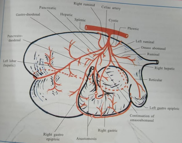
Celiac
artery
Origin- It is short but thick vessel arises from the abdominal aorta just after its entry into the abdominal cavity.
It is related to the pancreas and rumen at the left and right crus of diaphragm and caudal venacava at the right side.
Branches-
A. A. Caudal
phrenic and cranial adrenal arteries
B. B. Splenic
artery
C. C. Right
ruminal artery
D. D. Left
ruminal artery
E. E. Hepatic
artery
a.
Pancreatic
branch- pancreas
b.
Cystic
branch- gallbladder
c.
Right
hepatic branch - right part of liver
d.
Left
hepatic branch- left part of liver
i. Right
gastric
e.
Gastro-duodenal
artery
i. Pancreato-duodenal-
pancreas and duodenum
ii. Right
gastro-epiploic artery- abomasum and omentum
F. F. Left
gastric artery (omaso-abomasal artery)
i. Left gastro-epiploic artery
2.
Cranial mesenteric
artery
It arises from the aorta behind the celiac artery and then descends ventrally between the left lobe of pancreas and the caudal venacava. Then it passes caudally with the portal vein and crosses the caudal surface of the transverse colon.
Branches;
A. Pancreatic branches and the caudal pancreatico-duodenal artery
B. Middle colic artery
C. Ileocolic artery
D. Collateral artery
E. Jejunal artery
F. Ileal artery
3.
Renal artery(Left and right)
Right renal artery arises from the ventral aspect of the abdominal aorta at the level of second lumbar vertebra and passes laterally and cranially across the dorsal face of the caudal venacava and then enter the hilus of the kidney.
Left renal artery arises somewhat more caudally than the right renal artery and passes cranially and ventrally to reach the hilus of the kidney.
4.
Ovarian/testicular
artery
In male, the testicular artery arises from the ventral surface of the abdominal aorta close to the origin of the caudal mesenteric artery. After its origin it passes caudally in the sublumbar region and then ventrally, somewhat lateral to the pelvic inlet and descends towards the deep inguinal ring where it becomes a part of spermatic cord. It mainly supplies to the testicle, epididymis, ductus deferens and vaginal tunic.
In female, the ovarian artery originates in a same manner, but sometimes it arises from the external iliac artery. It is usually flexuous and following the free border of the broad ligament. It divides into small branches for uterine tube and uterine horn and reaches the ovary by the way of mesovarium.
5.
Lumbar artery
The lumbar arteries are six pairs in numbers, of which four or five pairs arise from the dorsal aspect of abdominal aorta. Usually the last pair, or sometimes also the fifth pair, arise from the internal iliac or iliolumbar artery. Their course and pattern of distribution are almost similar to that of intercostal arteries.
6. Caudal mesenteric artery
It arises from the abdominal aorta at the level of fifth lumbar vertebra and divides into a cranial colic branch and a caudal rectal branch to supply the colon and rectum.
Facebook Veterinary group


Post a Comment