Anjani Mishra
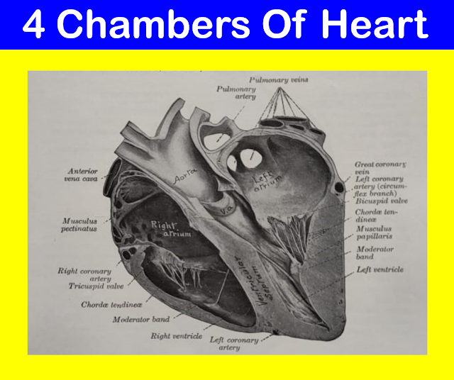
4 Chambers of the heart
Right
atrium
- Thin walled muscular chamber, smaller than the left atrium, receives venous blood from the body.
- It is placed at the anterior portion of the base of the heart. It is related to the left atrium on the left side and right ventricle ventrally.
- It possesses the sinus venosus, the main cavity and an appendix or auricle. Auricle is a small conical pouch forming a portion of the right and left atria of the heart. Each projects from the upper anterior portion of each atrium.
- The wall of atrium, particularly the
appendix presents some vertical muscular ridges and look like a comb and hence
named as pectinate muscles.
Opening
of right atrium:
- Cranial venacava:- It opens at the level of 4th rib.
- Caudal venacava:- It opens at the level of 5th rib.
- Vena hemi azygos:- It opens below or ventral to the opening of caudal venacava.
- Coronary sinus:- It opens very close to the vena hemi azygos.
- Right atrio-ventricular orifice (tricuspid valve):- It opens into the right ventricle.
- Foramen ovale (fetal circulation):- It opens into the left atrium through interatrial septum wall. Immediately after birth, it is closed and marks a small depression known as fossa ovale.
Vena hemi azygos- It is a single vein that drains blood from the dorsal wall of the left side of the thoracic and abdominal cavities and opens independently to the heart.

Fig: Vena hemi azygos
Left
atrium
- Thin walled muscular chamber, larger than the right atrium, receives oxygenated blood through pulmonary veins which open on its dorsal wall of the sinus venosus.
- It occupies the left caudal part of the base of the heart. It is related to the right atrium on the right side and left ventricle ventrally.
- The appendix is little larger and the pectinate muscles are less in number.
- The main cavity, sinus venosus is
comparatively larger than right atrium.
Opening
of left atrium:
- Four
pulmonary veins:- two from each lung
- Left
atrio-ventricular orifice (bicuspid/mitral/semilunar valve):- It opens into the
left ventricle.
- Foramen ovale:- The fossa ovalis is in the form of a faint depression.
Right
ventricle
- Thick walled conical chamber, larger than the right and left atrium, receives blood from the right atrium through right atrio-ventricular orifice, guarded by tricuspid valve and discharged it through the conus arteriosus into the pulmonary trunk.
- The conus arteriosus is placed at the left side and the right atrio-ventricular orifice is placed at the right side of the base of the ventricle.
- The tricuspid valve and the conus arteriosus is guarded by a thick ridge, the crista supraventricularis.
- It forms the right and cranial part of the heart.
- The wall of ventricle (except conus arteriosus)
bears muscular ridges and bands termed trabeculae carneae. It is of three
types;
a. Ridges
or columns- chordae tendineae
b. Musculi
papillares- papillary muscle
c. Muscular
band- moderator band- one in number, sometimes more than one, extends from the
lateral wall to the interventricular septum across the cavity of the ventricle.
Left
ventricle
- The wall of this chamber is thickest of all the compartments of the heart except apex. It receives blood from the left atrium through left atrio-ventricular orifice, guarded by bicuspid valve and discharged it through the aortic sinus into the ascending aorta.
- The aortic sinus is placed at the right side and the left atrio-ventricular orifice is placed at the left side of the base of the ventricle.
- It forms the left and caudal part of the heart and extends from the transverse groove to the apex.
- The trabeculae carneae are similar to
those of right ventricle. Moderator bands are usually two in number but thinner
than that of right ventricle.
Os-
cordis- found in sheep/ox
Ox-
two bones, ossa cordis, develop in the aortic fibrous ring.
- The right one is in apposition with the atrio-ventricular rings, and is irregularly triangular in shape.
- The left one is smaller and is inconstant. Its concave right border gives attachment to the left semilunar cusp of the aortic valve.
Sheep-
small os-cordis is single.
Pulmonary trunk
- This is a very short artery leaves the heart at the conus arteriosus at the base of the right ventricle between the appendices of right and left atria.
- It courses dorsally and caudally along the left side of the aorta, reaches the caudoventral aspect of descending aorta and divides into left and right branches.
- A little before its bifurcation into left and right pulmonary arteries, the common pulmonary trunk is connected to the descending aorta by a fibrous cord known as ligamentum arteriosum, the remnant of the ductus arteriosum of fetal life.
- The right pulmonary artery is longer and broader than the left. It passes caudally under the bifurcation of trachea and above the right atrium, reaches the hilus of right lung and enters below the right bronchus.
- The left pulmonary is short, passes caudally and enters the hilum of the left lung below the left bronchus.
Aorta
- Just at the beginning, i.e., close to the semilunar valves, the wall of the aorta is dilated in the form of a bulbus swelling, known as aortic sinus or bulbus aorticus. The left and right coronary arteries originate from the corresponding sides of the bulbus aorticus.
- The great arterial trunk originates at the base of the left ventricle and ascends between the pulmonary artery on the left and right atrium on the right. This beginning portion is called ascending aorta.
- The ascending aorta inclines backward after forming a sharp curve, the aortic arch. From the arch the vessel continues caudally along the dorsal aspect of the thoracic,abdominal and pelvic cavities as descending aorta.
Blood
supply to the heart
(Left and right coronary arteries)
Left
coronary artery
- It originates from the left side of the aortic sinus at the level of semilunar valves. It is larger than the right coronary artery.
- It passes between the left atrium and the pulmonary artery, reaches the atrioventricular groove and divides into descending and circumflex branches.
- The descending branch goes down along the left longitudinal groove and reaches the apical notch and anastomoses with the branches of the right coronary artery.
- The circumflex branch winds round the transverse groove caudally and reaches the right longitudinal groove and descends along it.
Right
coronary artery
- It is smaller and arises from the right side of the aortic sinus at the same level.
- It travels along the right part of the transverse groove and divides into a descending and a caudal branch.
- The descending branch travels along the right longitudinal groove and reaches the apical notch and anastomoses with the branches of the left coronary artery.
- Both the left and right descending branches supply to the interventricular septum.
Coronary vein
- A number of veins drains the blood from the walls of the heart. Many small veins return blood from the wall of the right side of the heart and opens at the vena azygos.
- The great cardiac vein receives the blood from the wall of both the ventricles and left atrium and opens into the coronary sinus.
- The middle coronary vein receives the blood from the region of interventricular septal wall.
Vasa vasorum
- The wall of the blood vessels are supplied with blood by numerous small arteries, called vasa vasorum. These arteries arises from the branches of artery which they supply or from adjacent arteries.
If you have any questions you can ask me on :
mishravetanatomy@gmail.com
Facebook Veterinary group
Facebook Veterinary group
Facebook - Anjani Mishra
Website: mishravetanatomy.blogspot.com


Post a Comment