Written By Anjani Mishra
Myology is the division of systematic anatomy which deals with the form and structure of muscles.
Muscle is an organ formed by muscular tissues composed of specialized cells which have the properties of contractility and conductivity to produce movement within certain organs and the body as a whole.
Muscle fiber:
The structural and functional unit of muscular tissue is a muscle fiber. Their arrangement suggests that they may be called fibers instead of cells, because muscle cells are much longer than their width, therefore muscle cells are often called muscle fibers.
It is a single cell in skeletal and smooth muscle, but in cardiac muscle, it is a collection of few cells.
These are cylindrical cells about 1mm to 4cm long in short muscle and upto
30cm in sartorius muscle. They vary in diameter from 0.01mm to 0.1mm or more.
A. An entire skeletal muscle enclosed within a dense connective tissue layer called epimysium
B. Each fascicle of muscle fibers is enclosed within a thin septa of C.T. called perimysium
C. Individual muscle fibers is surrounded by a more delicate C.T. called endomysium.
Myofibrils:
Each muscle fiber has bundles of thread-like contractile fibrils called myofibrils, made of actin and myosin myofilaments, which are actually protein molecules.
Fascia:
· A connective tissue membrane composed of bundles of white fibrous tissue,
separating muscles from each other and binding them into position are called
fascia.
· A more loosely packed layer next to the skin is the superficial fascia.
· It also unites the skin with underlying tissue, permitting the free
movement of the skin.
· A definite layer covering(investing) the groups of muscles and sending intermuscular septa is the deep fascia.
Aponeurosis:
· A flat fibrous sheet of connective tissue that serves to attach muscle to
muscle or other tissues. Sometimes, it may serves as a fascia.
· When fascia gives origin or insertion to muscle, it may appear as a
thickened sheet.
· An specially thick sheet of connective tissue acting in that capacity is
called an aponeurosis.
Tendon:
- A thick sheet of connective tissue composed of bundles of white fibers arranged in dense manner.
- It serves to attach the end of a muscle to the bone.
· Ligaments are strong bands or membranes usually composed of white fibrous
tissue which bind the bones together.
- They are pliable but practically inelastic. In few cases, however, eg; nuchal ligament, they are composed of elastic tissue.
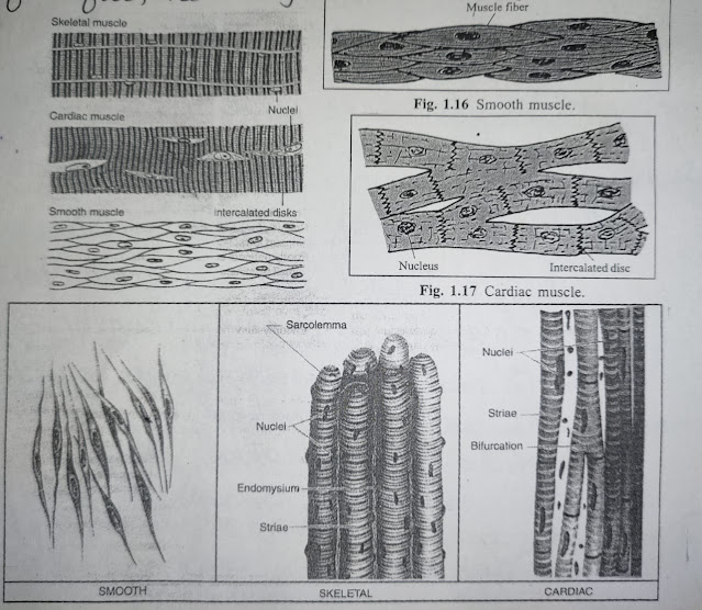
- Skeletal muscle - Voluntary, Striated
- Smooth muscle - Involuntary, non-striated
- Cardiac muscle - Involuntary, striated
Skeletal muscle:
It consists of bundles of very long, cylindrical,
multinucleated cells. Their contraction process is quick, forceful and usually
under voluntary control.
Smooth muscle:
It consists of masses of spindle-shaped cells in
the wall of visceral organs and blood vessels. Their contraction process is
slow, involuntary.
Cardiac muscle:
Types of skeletal muscle fibers
1.
Parallel muscles
The
muscle fibers are arranged parallel to the line of pull. The fibers are long,
but their numbers are relatively few.
Subtypes
of parallel muscles;
a. Strap- eg; sartorius, sternohyoid etc.
b. Strap with tendinous intersection- eg; rectus
abdominis etc.
c. Quadrilateral- eg; quadratus lumborum, quadratus
femoris etc.
d. Fusiform- eg; biceps brachii, digastricus etc.
2. Oblique muscles
In this
type of muscles, the muscle fibers are oblique to the line of pull.
Subtypes
of oblique muscles;
a.
Pennate
Muscle
fibers are shorter, but more in number, having feather like appearance. The
fleshy fibers are arranged obliquely in the long axis of the muscle and in the
direction of the muscle pull.
Subtypes
of pennate muscles;
i.
Unipennate- all fleshy fibers slope into one side of the tendon, which is formed along one margin of the muscle. This gives the appearance of half of feather. Eg; extensor digitorum longus, peroneus tertius etc.
ii. Bipennate- the tendon is formed in the central axis of the muscle and the
muscle fibers slope into the two sides of the central tendon, like a whole
feather. Eg; rectus femoris etc.
iii. Multipennate- looks like many feathers, where a series of bipennate
muscles are arranged in side by side having a tendon centrally which extends
from its insertion to its origin. Eg; deltoid, sub-scapularis etc.
b. Triangular
Muscle
fibers converge on an apical tendon, i.e. originated from the base and inserted
into the apex which is triangular in shape. Eg; temporalis, adductor longus
etc.
3. Spiral muscle
In this
muscles, the fibers are twisted in arrangements close to their insertion. Eg;
latissimus dorsi, trapezius, pectoralis etc.
4.
Cruciate muscle
What do
you mean by origin and insertion of a muscle ?
Origin- generally proximal, fleshy, fixed end is called
origin.
mishravetanatomy@gmail.com
Facebook Veterinary group
Facebook - Anjani Mishra


Post a Comment