
GENERAL OSTEOLOGY
(Skeletal System)
OSTEOLOGY: is the division of systematic anatomy which deals with description of the skeleton (bones and cartilage).
(Skeletal System)
Written By Anjani Mishra
SKELETON: The term skeleton is applied to the
frame work of hard structures which supports and protects the soft tissues of
animals. In the descriptive Anatomy of higher animals, it is usually restricted
to the bones and cartilages, although the ligaments which bind these together
might well be included.


Classification
Skeleton
Skeleton
A. Appendicular B. Axial C. Visceral
|
Include the bones of limbs; forelimb or thoracic limb, hindlimb
or pelvic limb |
Includes the bones of head, vertebral column, ribs and sternum |
Includes certain bones developed in the substance of some of the
viscera or soft organs |
Skeletal formula – Express the number of total bones in the skeleton.
Skeleton formula of domestic animals and fowl:
|
Region |
Cattle |
Horse |
Dog |
Pig |
Fowl |
|
Vertebral column |
50 |
54 |
52 |
52 |
41 |
|
Ribs and sternum |
27 |
37 |
27 |
31 |
15 |
|
Forelimb |
50 |
40 |
88 |
76 |
28 |
|
Hindlimb |
50 |
40 |
88 |
76 |
42 |
|
Skull |
34 |
34 |
34 |
30 |
40 |
|
Viscreal bones |
2 |
-- |
1 |
1 |
1 |
|
Total |
213 |
205 |
290 |
266 |
167 |
Bone: is one of
the hardest connective tissue of the body which is suited for its supportive
and protective function in the skeleton.
Classification:
The bones (ossa) are commonly divided into four classes according to their
shape and function.This classification is not entirely satisfactory; some
bones, e.g., the ribs, are not clearly provided for, and other might be
variously placed.
- Long bones (ossa longa): Long bones are typically of elongated cylindrical form with enlarged extrimities. They occur in the limbs, where they act as supporting columns and as levers. The cylindrical part, termed the shaft or body (corpus), is tubular and encloses the medullary cavity (cavum medullare), which contains the medulla or marrow.
- Flat bones (ossa plana): Flat bones are expanded in two directions. They furnish sufficient area for the attachment of muscles and afford protection to the organs which they cover. e.g., scapula and many bones of the skull. Flat bones consists of two layers of compact bone with intervening spongy bone and marrow. The spongy layer in the bones of the skull is termed diploe.
- Short bones (ossa brevia): Short bones present somewhat similar dimensions in length, breadth and thickness. Their chief function appears to be that of diffusing concussion. e.g., carpus, tarsus and sesamoid bones. They diminish friction or change the direction of tendons or increase leverage to muscles and tendons.
- IRREGULAR BONES: This group include the bones of irregular shape and they are median and unpaired. Their functions are various and not so clearly specialized as those of the preceding classes. e.g., vertebrae and the bones of the cranial base.
Functions:
·
Supports and protects the soft tissues of the body.
·
Plays vital role in the motility of
the animal.
·
Important source of essential
minerals like calcium and phosphorus.
·
Important source of blood cells like
RBC, WBC, & platelets.
·
Provide body shape and bears the weight
of animal body.
Structure of bone
The
architecture of bone can be studied best by means of longitudinal and
transverse sections of specimens which have been macerated so as to remove most
of the organic matter. These show that the bone consists of an external shell
of dense compact substance, within which is the more loosely arranged spongy
substance. In typical long bones the shaft is hollowed to form the medullary
cavity.
Major
constituents of bone
- Periosteum
- Compact bone
- Spongy bone
- Endosteum
- The marrow
1) Periosteum
– is the membrane which invest the
outer surface of bone, except where it is covered with cartilage. It is a layer
of specialized connective tissue. A periosteum layer is lacking on those areas
of the epiphyses of long bones that are covered with articular cartilage. The
periosteum consists of an outer protective fibrous layer and an inner cellular
osteogenic layer. During active growth the osteogenic layer is well developed,
but later it becomes much reduced. The fibrous layer varies much in thickness,
being, in general, thickest in exposed situations. The adhesion of the
periosteum to the bone also differs greatly in various places; it is usually
very thin and easily detached where it is thickly Covered with muscular tissue
which has little or no attachment.
2) Compact bone –
differs greatly in thickness in various situations, in conformity with the
stresses and strains to which the bone is subjected. In the long bones it is
thickest in or near the middle part of the shaft and thins out toward the
extremities. It is specially dense and smooth on joint surface. Circumscribed
thickenings are found at points which are subject to special pressure or
traction.
3) spongy bone
– consists of delicate bony plates and spicules which runs in various
directions and intercrosses. These are definitely arranged with regard to
mechanical requirements, so that systems of pressure and tension plates can be
recognized in conformity with the lines of pressure and the pull of tendons and
ligaments respectively.The intervals between the plates are occupied lay marrow
and are termed marrow spaces.The spongy substance forms the bulk of short bones
and of the extremities of long bones; in the latter it is not confined to the
ends but extends a variable distance along the shaft also.


Some
bones contain air spaces within the compact substance instead of spongy bone
and marrow and hence, are called pneumatic bones. The cavities are termed
sinuses and are lined with mucous membrane; they communicate indirectly with
the external air. In certain situations the two compact layers of flat bones
are not separated by spongy bone, but fuse with each other; in some case of
this kind the bone is so thin as to be translucent, or it may undergo
absorption, producing on actual deficiency. The
flat bones of the cranial vault and sides are composed of an outer layer of ordinary
compact substance, the lamina externa, an inner layer of very dense bone, the
lamina interna or tabula vitrea end between these a variable amount of spongy
bone, here termed diploe.
4 ) endosteum
– the endosteum is a thin fibrous membrane which lines the medullary cavity
and
the larger haversian canals (nutrient canal of bone).
5) The
marrow – occupies the interstices of the
spongy bone and the medully cavity of the long bones. There are two
varieties in the adult - red and yellow. In the young subject there is only red
narrow but later this is replaced in the medullay cavity by yellow marrow. The
red marrow contains several types of characteristics cells and is a blood
forming substance, where as the yellow is practically ordinary adipose tissue.
Yellow marrow is formed by regressive changes in red marrow, including fatty
infiltration and degeneration of the characteristics cells; thus we find transitional
forms or stages in the process. In aged or badly nourished subjects the narrow
may under go gelatinous degeneration, resulting in the formation of gelatinous
marrow. Red narrow persists in the sternum through out the life and thus, this
is a convenient place for examination and aspiration.
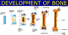
Development
and Growth of bone
The primitive embryonal skeleton
consists of cartilage and fibrous tissue, in which the bones develop. The
process is termed ossification or osteogenesis, and is effected essentially by
bone-producing cells, called osteoblasts. It is customary, therefore, to
designate as membrane bones those which are developed in fibrous tissue, and as cartilage bones those
which are preformed in cartilage. The principal membrane bones are those of the
roof and sides of the cranium and most of the bones of the face. The cartilage
bones comprise, therefore, most of the skeleton.
Correspondingly we distinguish intramembranous and endochondral ossification.
In intramembranous ossification the process begins at a definite center of
ossification (Punctum ossificationis),where the osteoblasts surround
themselves with a deposit of bone. The process extends from this center to the
periphery of the future bone, thus producing a network of bony trabeculae. The
trabeculae rapidly thicken and coalesce, forming a bony plate which is
separated from the adjacent bones by persistent fibrous tissues.
The superficial part of the original
tissue becomes periosteum, and on the deep face of this successive layers of
periosteal bone are formed by osteoblasts until the bone attains its definitive
thickness. Increase in circumference takes place by ossification of the
surrounding fibrous tissue, which continues to grow until the bone has reached
its definitive size. In endochondral ossification the process is fundamentally
the same, but not quite so simple. Osteoblasts emigrate from the deep face of
the perichondrium or primitive periosteum into the cartilage and cause
calcification of the matrix or ground substance of the latter. Vessels extend
into the calcifying area, the cartilage cells shrink and disappear, forming
primary marrow cavities which are occupied by processes of the osteogenic
tissue. There is thus formed a sort of scaffolding of calcareous trabeculae on
which the bone is constructed by the osteoblasts. At the same time perichondral
bone is formed by the osteoblasts of the primitive periosteum. The calcified
cartilage is broken down and absorbed through the agency of large cells called
osteoclasts, and is replaced by bone deposited by the osteoblasts. The
osteoclasts also cause absorption of the primitive bone, producing the marrow
cavities; thus in the case of the long bones the primitive central spongy bone
is largely absorbed to form the medullary cavity of the shaft, and persists
chiefly in the extremities. Destruction of the central part and formation of
subperiosteal bone continue until the shaft of the bone has completed its
growth.
Chemical
and physical properties of bone
Dried
bone consists of organic and inorganic matter in the ratio of
approximately 1:2. The animal matter gives toughness and elasticity, the
mineral matter gives hardness to the bone tissue. Removal of the organic matter
by heat does not change the general form of a bone, but reduces the weight by
about one third and makes the bone very fragile. Conversely, decalcification,
while not affecting the form and size of the bone, makes it soft and pliable.
The organic matter (ossein) when boiled yields gelatin. The organic portion of
bone consists chiefly of a protein, called bone collagen or ossein. Bone itself
is a highly specialized form of C.T. that is hard and white and contains cells
peculiar to it.
The
hardness of bone is due to the deposition of mineral salts within the soft
organic matrix. In addition to containing water bone consists of two main
components;
1. The organic framework, and
2. The inorganic mineral salts (bone ash)
Between the collagenous fibres a
fluid is found, resembling tissue fluid, an amorphous ground substance(mucopolysaccharides, hyaluronic acid, condroitin sulphate, keratin, and electrolytes). The
following table represents the composition in 100 parts of ox bone of average
quality:
Gelatin -----------------------------------------------------
33.30
Calcium phosphate
--------------------------------------- 57.35
Calcium carbonate ----------------------------------------
3.85
Magesium phosphate
------------------------------------- 2.05
Carbonate and chloride of sodium ----------------------
3.45
100.00
Fresh dead bone has a yellowish
white color; when macerated or boiled and bleached, it is white. The specific
gravity of fresh campact bone is about 1.9. It is very hard and resistent to
pressure. Its compressive strength is about 20,000 pounds per square inch, and
its tensile strength averages 15,000 pounds per square inch.
composition of bone
The hard extremely dense connective
tissue that forms the skeleton of the body. It is composed of a matrix of
collagen fibers impregnated with bone salts (chiefly calcium carbonate and
calcium phosphate).
- Collagen fibers
by weight:
1/3 of bone
by volume:
1/2 of bone
- hydroxyapatite crystals
(ca)10(PO4)6(OH)2
95% solid (Vs. Water)
65% mineral; 35% Organic
Organic matter:
The growth and development of the
bone is possible due to the organic matter contained in it. Organic matters are
present in the bone in the form of membrane, cartilage, marrow, vessels, nerves
and included fluids.
Inorganic matter:
Inorganic matters are present in the
from of salts, deposited during the process of ossification of bone. The
strength and hardness of a bone is due to the inorganic salt constituents.
If you have any questions you can ask me on :
mishravetanatomy@gmail.com
Facebook Veterinary group link - https://www.facebook.com/groups/1287264324797711/
Twitter - @MishraVet
Facebook - Anjani Mishra
mishravetanatomy@gmail.com
Facebook Veterinary group link - https://www.facebook.com/groups/1287264324797711/
Twitter - @MishraVet
Facebook - Anjani Mishra
Website: mishravetanatomy.blogspot.com
Post a Comment