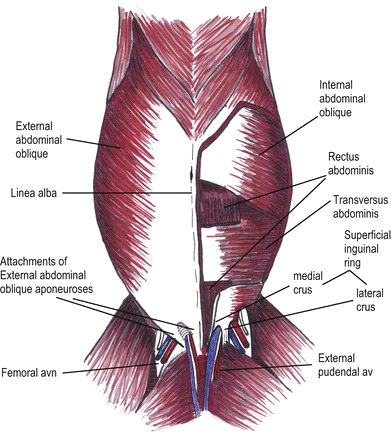
Muscles of abdomen
- The abdominal muscles form much of the lateral and all of the ventral abdominal wall and are named according to the direction of their fibers.
- Contraction of the abdominal muscles places pressure on the abdominal viscera and thus aids in micturition, defecation, parturition, forced expiration and coughing.
- The contraction of the abdominal muscles and the diaphragm on the viscera is known as the “abdominal press.”
Muscles of abdomen are as follows;
- The obliquus externus abdominis muscle
- The obliquus internus abdominis muscle
- The rectus abdominis muscle
- The transversus abdominis muscle
- The cremaster externus muscle(male)
1. 1. The Obliquus Externus Abdominis Muscle
It is the most extensive of the abdominal muscles. It is a broad
sheath, irregularly triangular in shape, whose fibers are directed ventrad and
caudad.
Origin: The caudal border and lateral surface of the
last eight(8) ribs.
Insertion: Linea-alba, prepubic tendon and coxal tuber by aponeurotic tissue.
Action:
Compression of the abdominal viscera, so aids in abdominal press.
Blood supply: Lumbar and dorsal intercostal & circumflex
iliac artery.
Nerve supply: Intercostal and lumbar nerves.
2. 2. The Obliquus Internus Abdominis
Muscle
It
is situated beneath the obliquus externus abdominal muscle. Its fibers are
directed ventrad, craniad and mediad. It is triangular in shape with the base
caudad.
Origin: The coxal tuber and the deep lumbar fascia.
Insertion: Linea-alba, prepubic tendon and the caudal border of the last rib.
Action:
Compression and support the abdominal viscera.
Blood supply: Lumbar and dorsal intercostal & circumflex
iliac artery.
Nerve supply: Intercostal and lumbar
nerves.
3. 3. The Rectus Abdominis Muscle
It is confined to the ventral abdominal wall. It
extends from the sternum to the pubis. It lies or arises on the lateral border
of the sternum.
Origin: The ventral and lateral surface of the sternum
as far craniad as the third(3rd ) or fourth (4th )costal
cartilage.
Insertion: The prepubic tendon, and thus, indirectly to the pectin ossis pubis and symphysial ligament.
Action:
Aids in abdominal press.
Blood supply: Lumbar and dorsal intercostal & circumflex
iliac artery.
Nerve supply: Intercostal and lumbar
nerves.
4. 4. The Transversus
Abdominis Muscle
This
muscle layer is the deepest in the abdominal wall and forms the wall, particularly in the flank region.
Origin: The deep lumbar fascia, the medial surface of
the false rib, the transverse fascia and thus indirectly to the first five
lumbar transverse processes
Insertion: The linea alba.
Action:
To retract the ribs and compress the abdominal viscera.
Blood supply: Lumbar and dorsal intercostal & circumflex
iliac artery.
Nerve supply: Intercostal and lumbar nerves.
5. 5. The Cremaster Externus Muscle(in male only)
·
This muscle may be regarded as a detached
portion of the obliquus internus abdominis muscle which separates as a slip of
fleshy tissue to enter the inguinal canal.
·
In rumunants, it is well developed and
almost completely encloses the vaginal tunic to the neck of the scrotum.
·
It is inserted at about the level of the
head end of the testicle, and arises by a thin aponeurosis, which is succeeded
by a flat muscular belly.
· It descends through the inguinal canal on the caudo-lateral surface of the vaginal tunic to which it is very loosely attached.
Insertion: The internal spermatic fascia.
Linea-alba
·
It is a white fibrous raphe that extends
from the xyphoid cartilage to the pre-pubic tendon.
· It results from the union of the aponeurosis of the oblique and transverse abdominal muscles.
Pre-pubic
tendon
·
It is essentially the tendon of insertion
of the two rectus abdominis muscle.
·
The ventral surface of the pre-pubic
tendon also provides origin for the symphysial tendon (well developed in
female), which furnishes an attachment to the obliquus externus abdominis,
gracilis and pectineus.
·
The pre-pubic tendon is attached to the
cranial borders of the pubic bones, including ilio-pubic eminence. It has the
form of very strong, thick band with concave lateral borders which, in turn,
form the medial boundry of the superficial inguinal ring.
·
Rupture of the pre-pubic tendon occasionally
occurs in the ox, particularly during gestation, resulting in the relaxation of
the abdominal musculature and a concave appearance to the dorsum.
Inguinal canal
- The term inguinal canal or space is applied to an oblique passage through the caudal part of the abdominal wall.
- The paired canals lie on either side of the pre-pubic tendon.
- The canal begins at the deep inguinal ring and extends obliquely ventro-medially to end at the superficial inguinal ring.
- In the male, this slitlike passage or space between the abdominal muscles contains the spermatic cord, vaginal tunic, cremaster muscle, external pudendal artery and, inconstantly, a small satellite vein, as well as the inguinal lymph vessels and the ilioinguinal and genitofemoral nerves.
- In the female, it contains the external pudendal vessels and ilioinguinal and genitofemoral nerves.
- The cranial wall of the canal is formed by the caudal part of the obliquus internus abdominis. The caudo-lateral wall is formed by a portion of the aponeurosis of the obliquus externus abdominis.
- The deep inguinal ring is the abdominal opening of the canal. It is bounded cranially by the margin of the obliquus internus abdominis and caudally by the inguinal ligament. Medially, it is related to the concave lateral border of the pre-pubic tendon. The lateral limit of the ring is formed by the attachment of the obliquus internus abdominis to the inguinal ligament.
- The deep inguinal ring is directed from the edge of the pre-pubic tendon toward the coxal tuber in a dorsal and lateral direction. It has a length of approximately 15 cm in the ox and 2.5 cm in goat.
- The superficial inguinal ring is well defined slit in the aponeurosis of the obliquus externus abdominis lateral to the pre-pubic tendon. Its boundaries are the medial and lateral crura.
- The direction of the axis of the superficial inguinal ring is ventrad, craniad and laterad. The medial angle of the two rings are separated only by a distance equal to the thickness of the pre-pubic tendon, whereas the lateral angles are approximately 15-17.5 cm apart in the ox and a correspondingly lesser distance in small ruminants.
- The medial angle of the superficial inguinal ring is well defined and is distinctly palpable at the side of the pre-pubic tendon.
Facebook Veterinary group


Post a Comment