
URINARY SYSTEM
Written By Anjani Mishra

Kidneys
The kidneys are paired organs situated
retro-peritonially on the posterior wall of the abdominal cavity.

Fig: Urinary system of dog

Fig: Urinary system of human
Structure
Capsule- the kidneys are covered by a connective tissue
capsule consists of a collagen fiber network with small amount of elastic
fibers.
A longitudinal Sections of kidney present;
1) A peripheral darker zone- the cortex,
2) An inner lighter zone- the medulla.
The cortex consists of renal corpuscles, proximal
and distal convoluted tubules. The medulla consists of loop of henle's (both descending & ascending) and straight tubule which converge towards the
apex called medullary rays.
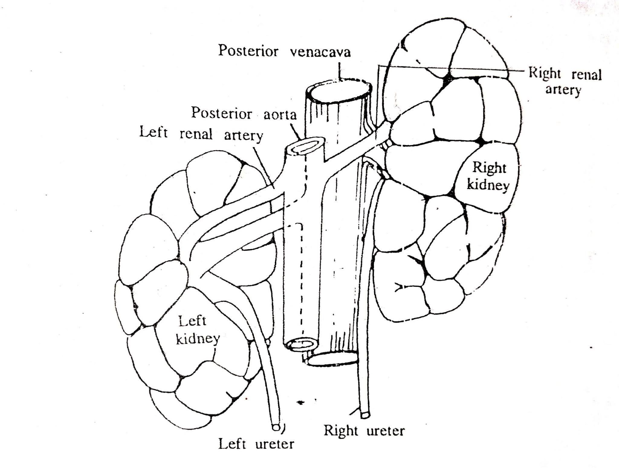
Fig: Schematic diagram of the
kidneys of cow

Fig: Section of kidney
of ox
 Fig: Schematic diagram of uriniferous tubule
Fig: Kidney, cortex and portion of medulla, dog
3. Capsule, 7. Medulla, 8. Pars convoluta, 9. Pars radiata, 11. Renal corpuscle
Uriniferous
tubules
The functional unit of the kidney is the
uriniferous tubules, which is highly convoluted structure involved in urine
formation. Each uriniferous tubules consists of a secretory part, called the
nephron, and a collecting tubule.
Nephrons
Nephrons are the basic functional subunits
of the kidney. Each nephron consists of renal corpuscle, proximal convoluted
tubule, thick and thin limbs of Henle's Loop, and distal convoluted tubule.
Renal
corpuscle
It consists of glomerulus and the Bowman's
capsule. It is oval to round structure about 200mm in diameter, where filtration of blood
occurs.
Glomerulus
- It is a tuft of capillaries that extends into the Bowman's capsule. It is formed by the branches of the afferent arteriole.
- It is lined with glomerular endothelial cells that form the inner layer of the capillary wall. They have large fenestrae (gap).
- A special type of cell, called mesangial cells lie between the capillary loops. These cells can contract and there by decreasing the surface area available for filtration.

2. Capsular epithelium, 5. Distal convoluted tubule, 6. Glomerular epithelium,
10. Proximal convoluted tubule, 14. Urinary space
Bowman's
(renal) capsule
- It is a double walled epithelial capsule, composed of parietal and visceral layer. In between the parietal and visceral layer, there is a narrow space called Bowman's Space (or urinary space).
- The parietal layer is composed of simple squamous epithelium, forming the outer limit of the renal corpuscle.
- The visceral layer is composed of modified simple Squamous epithelium, the podocytes, that envelops the capillaries of the glomerulus.
- Podocytes have a cell body from which arises several primary processes. Each primary process gives rise to numerous secondary processes, called, pedicels. Pedicels that embrace the capillaries of the glomerulus.
Proximal
Convoluted tubule
- An epithelial tube that begins at the urinary pole of the renal corpuscle.
- It is lined with simple cuboidal to columnar epithelium
- The living cells have abundant long microvilli. Together they form brush border that partly obscures the lumen and increase the surface area available for absorption.
Henle's loop
It is U-shaped epithelial tube consisting
of a thick descending limb, a thin descending limb, a thin ascending limb, and
a thick ascending limb.
Descending loop of henle (Thick)
- It is lined by sim. Cuboidal epithelium that has a prominent brush border.
- It is also known as the straight portion of the proximal tubule.
Descending
loop of henle (thin)
- It is lined with simple Squamous epithelium.
- They have few short microvilli.
Ascending
loop of henle (thin)
- Same as above
Ascending
loop of henle (thick)
- It is lined by simple Cuboidal epithelium cells that posses a few microvilli.
- It is also known as the straight portion of the distal tubule.

Fig: Kidney, cortex, dog
1. Adipose tissue, 3. Capsule, 8. Pars convoluta, 9. Pars radiata, 11. Renal corpuscle
Distal convoluted tubule
- It is much shorter and has a wider lumen than the proximal convoluted tubule.
- It is lined by simple Cuboidal epithelium cells.
- They do not contain brush border.
- It establishes contact with the vascular pole of renal corpuscle. At this point of close contact, the wall of the distal tubule is modified, forming macula densa, which is the part of juxtaglomerular apparatus(JGA).

Juxtaglomerular apparatus(JGA)
It is located at the vascular pole of the renal corpuscle. It consists of following structures;
- Macula densa of the distal tubule,
- Juxtaglomerular cells of afferent and occassionally efferent glomerular arterioles, and
- Juxtaglomerular mesangial cells, also known as polkissen/lacis cell
Collecting tubules
- They have the segments in both the cortex and medulla.
- It is lined by simple Cuboidal epithelium cells (cortex)
- They don not have brush border.
- Medullary collecting tubules are lined by simple columnar cells.
- They have larger lumen than the convoluted tubules.
(Note:Figure for practical note book)

Ureter
The ureter conveys urine from the kidney to the urinary bladder.
Structure
The wall
of ureter has 3 layers;
1 1. Mucosa
a. Lamina
epithelia
b. Lamina
propria
2 2. Muscularis
externa
3. Adventitia
3. Adventitia

Fig: Ureter, x.s., cat
1. Adventitia, 2. Lamina propria, 4. Muscularis, 7. Transitional epithelium
1. Mucosa
a. Lamina epithelia- It is lined with
transitional epithelium with a wide variety of cell forms. In the distended
ureter all cell appear in section to be narrow spindle.
b. Lamina propria- It consists mainly of
collagen fiber, blood vessels, lymphatics. In horse, it contains tubulo-alveolar
mucous glands with wide lumina. The lumen has a star-shaped appearance, that is
due to the longitudinal folds of the mucosa.
Muscularis mucosa and sub-mucosa- absent
2. Mucularis externa
It consists of thin bundle and forms;
- An inner longitudinal,
- A middle circular, and
- A thin outer longitudinal layer, which may occasionally
be interrupted by collagenous bundles. Near the opening into the bladder, the
ureter contains only longitudinal muscle fiber.
3. Adventitia
External to the muscularis, a loose adventitia composed of collagen and reticular fiber contains the larger blood vessels and nerves
External to the muscularis, a loose adventitia composed of collagen and reticular fiber contains the larger blood vessels and nerves

2. Lamina propria, 3. Mucous gland, 7. Transitional epithelium
Urinary bladder

Fig: Urinary bladder, cow
2. Lamina propria, 4. Muscularis, 5. Muscularis mucosae, 6. Sub-mucosa,
7. Transitional epithelium
The wall of urinary bladder consists of ;
1.Mucosa
a. Lamina epithelia – lined with
transitional epithelium.
b. Lamina propria – the collagenous propria
contains few elastic fibers.
c. Lamina muscularis mucosa – there is
usually a thin layer of longitudinal muscle in between the
lamina propria and a
loose sub-mucosa.
2. Sub-mucosa
– Consists
of loose connective tissue
3. Muscularis
externa
– It consists of coarse bundle of fibers and are arranged in :
- An
inner longitudinal layer
- A middle circular (thickest layer)
- An outer longitudinal layer
The
middle circular layer forms the sphincter around the internal urethral orifice,
where as outer longitudinal may be modified or absent in some places.
4. Serosa/
adventitia
A serosa is present on the apex and body
of the bladder. At the neck, the muscularis is covered by an adventitia of the
same structure as ureter.

Fig: Urinary bladder, goat
1. Capillary, 5. Lamina propria, 9. Transitional epithelium
Urethra

Fig: Genital organs of bull


Fig: Genital organs of cow

Fig: Genital organs of bull
1. Ureter, 2. Prostate gland, 3. Seminal vesicle, 4. Urethral muscle, 5. Bulbourethral gland

Fig: Pelvic urethra

The male urethra
The urethra is divided into pelvic portion and
an extra-pelvic portion or penile portion.
1. Pelvic
urethra
Structure
The wall of penile urethra consist of following layers;
a. Lamina epithelia
– The pelvic urethra
is lined by a longitudinally folded mucosa which bears transitional epithelium in most of the domestic animals, but in case of dog, cat and buck it is lined with stratified columnar epithelium showing evidence of secretory activity. The surface of the epithelium is irregular.
b. Lamina propria
– The thin propria is collagenous and poorly vascularized.
Corpus cavernosum urethrae
– Next to the mucosa
lies the stratum vasculare or cavernosum, which is present in all species.
It is composed of dense plexus of veins and have the character of erectile tissue.
The trabeculae between the veins are rich in smooth musculature. The adjoining
glandular layer contains the prostatic tissue (or urethral glands).
c. Lamina muscularis
mucosa – The
next layer is a sheet of thin bundles of smooth muscles, followed by the striated
urethral muscle.
 Fig: Penile urethra, x.s., ram

2. Cavernosus space, 10. Tunica albuginea, 11. Urethra, lumen
2. Extra pelvic/ Penile urethra
Structure
- The penile urethra has fewer glands but more erectile tissue than the pelvic portion. The folded mucosa bears transitional epithelium, which changes at the urethral opening to stratified squamous epithelium on a papillary body.
- The lamina propria varies in thickenss. In the bull it has the character of lymphatic tissue, but in the stallion and boar, it contains the small scattered 'glands of Littre.'
- The cavernosus tissue adjoining the mucosa is composed of connective tissue trabeculae which are continuous peripherally with the tunica albuginea that limits the erectile tissue. The trabeculae contain many elastic fibers and blood vessels, and in the horse and bull, longitudinal smooth muscle fibers.
- Between the trabeculae there are spaces increasing in size towards the albuginea, which are lined with endothelium and usually filled with blood. Towards the mucosa these spaces pass into an extensive venous plexus which occupies the deeper layers of the mucosa.
- The cavernosus spaces receive their blood from these veins and from the capillaries of the trabecular system. Here the artery of the urethral bulb opens directly into the cavernosus sinuses.
If you have any questions you can ask me on :
anjanivetanatomy@gmail.com
Twitter - @MishraVet
Facebook - Anjani Mishra
anjanivetanatomy@gmail.com
Twitter - @MishraVet
Facebook - Anjani Mishra
Website: mishravetanatomy.blogspot.com
Post a Comment