
Accessory Organs of Digestive System

Written by Anjani Mishra
Accessory organs of digestive system are:
(Tongue, Teeth, Salivary gland, Pancreas, and Liver)

TONGUE
GROSS
- The tongue is a highly movable muscular organ covered by mucous membrane.
- It is attached to the floor of the mouth between the bodies of the mandible.
- It consists of three parts, namely; root(redix linguae), body(corpus linguae), and apex(apex linguae).
- Root and body are wide where as apex is free and pointed and has a rounded margin except fowl.
- It is mainly composed of mucous membrane, muscles, glands, blood vessels and nerves.
- It is important in the prehension, mastication, deglutition, voice and taste of food.
HISTOLOGY
- The epithelium is stratified squamous with varying thicknesses of the stratum corneum.
- It is thickest on the dorsal surface and thinnest on the ventral surface, where it is non-keratinized.
- The dorsal surface contains numerous papillae.
- These papillae differ in shape and named according to their morphologic characters.
- They either serve mechanical or gustatory (sense of taste) function.
- The filiform, conical, and lenticular papillae facilitate the movement of the ingesta within the oral cavity, where as fungiform, vallate, and foliate contain taste buds, which is responsible for mediation of the sense of taste.

Fig: Tongue, goat
4. Filiform papilla
5. Fungiform papilla
7. Skeletal muscle
8. Small papilla
1) The filiform papillae are the most numerous types. They
are slender, sharp- pointed structures that projected above the surface of the tongue
and are covered by keratinized stratified squamous epithelium with a thick
stratum corneum. They are supported by a high vascularized connective-tissue
core.
(2) The conical papillae and lenticular papillae are
characterized by cone shape and lentil-shape. respectively and are located on
the torus linguae of ruminant animals.
(3) The fungiform papillae are scattered among the filiform
papillae. The shape is suggested of a mushroom. The taste bud is located on the
upper surface and the lining epithelium is non-keratinized epithelium. The
connective tissue core is rich in blood vessels and nerves.
(4) The vallate papillae are large and flattened with gustatory
furrow. The taste buds are located on the lateral side of the papillae. These papillae are encircled by a wall, hence known as circumvallate.
(5) The foliate papillae are common in horse, dog, cat, pig, and rabbit
characterized by thin plate of elongated fold with gustatory furrow. The taste buds are
located in the epithelium of the sides of the fold.
Histology of the taste buds:

Fig: Taste bud
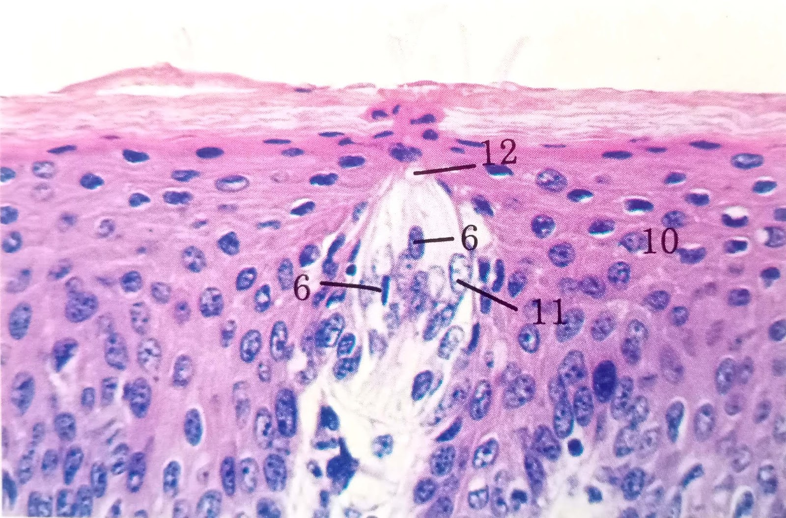
Fig: Tongue, horse
6. Sensory cell, nucleus
10. Stratum spinosum
11. Supporting cell, nucleus
12. Taste pore
- The taste buds are ellipsoid clusters of sensory taste cells in the stratified squamous epithelium of the fungiform, vallate and foliate papillae of the tongue.
- They also occur widely dispersed in the soft palate, epiglottis or other areas of mouth and pharynx.
- The taste buds are flask shaped structures with a base, wide body and neck which opens at the taste pore.
- The taste bud consists of spindle-shaped taste cells/gustatory cells at the centre and crescent shaped supporting cells at the peripheral part.
- Some of the cells have microvilli at their apex which is considered to be a supporting cells and the cells have no microvilli are the chemoreceptor cells responsible for taking taste.
- Tongue consists of both extrinsic and intrinsic muscles;
- The intrinsic striated lingual muscle mass composed of longitudinal, transverse, and vertical and are arranged obliquely, responsible for the movement of the tongue.
- The dorsal longitudinal muscle fibers are situated below the mucosa.
- Scattered among the muscle fibers and clusters of sero-mucous glands in the sub-mucosa are collectively called lingual glands.
- Lingual tonsils are small and numerous located at the base of tongue lined by stratified squamous epithelium.
- Lyssa is a fibro-elastic cord extends along the midline of the ventral surface of the tongue of dog composed of fibrous C.T., adipose tissue, striated muscle fibers, vessels and nerves. It is a characteristics feature of dog.
- In cat it is mainly composed of adipose tissue. In other animals it is composed of fibro-elastic tissue.

Fig: Tongue, musculature, l.s., cat
4. Filiform papilla
11. Muscle, longitudinal
11. Muscle, longitudinal
12. Muscle, Transverse
13. Muscle, verticle
Teeth
GROSS
- Grossly, the tooth consists of a root, which is contained within the alveolus.
- The exposed part is the crown, projecting from the alveolus.
- The intermediate part is the neck, which is sorrounded by the gum.
- In some domestic animals there is no constriction at the neck, and the enamel-covered body of the tooth extends far down into the alveolus.
- Histologically, five major constituents of the tooth has to be concider for description;
- It consists of three hard substances; enamel, dentin, and cementum, as well as
- Two soft substances; pulp cavity/dental pulp and the periodontal membrane.
- The enamel, is the hardest substances in the body which covers the crown and forms a thin layer over the surface of the tooth.
- It is almost devoid of organic matter and consists almost entirely of inorganic material.
- In addition to a small amount of cementing substances, it contains long, slender, hexagonal, calcified rods called enamel prisms.
- Each prism exhibits cross striation. The prism bundles run in spirals and waves to the outer enamel surface.
- The variation in the curves of the bundles gives the false impression that they cross.
- The elongated spaces filled with cementing material occur between the prism.

Fig: Dentinoenamel junction, dog
2. Ameloblasts, 10. Dentin, 11. Enamel, 16. Odontoblasts, 18. Pre-dentin, 19. Space artifact,
20. Stellate reticulum, 21. Stratum intermedium
- The dentin, is the main substance of the tooth which is similar in composition to bone, but differ structurally in that; it contains numerous fine parallel tubules called dentinal tubules.
- It is a peculiar tissue in which the cells, called odontoblasts, remain on the inner surface lining the pulp cavity and send long processes out through the calcified substance.
- It contains collagenous fibrils which run generally parallel to the surface in the crown, parallel to the occlusal surface.
- When decalcified, dentin resembles cartilage. It is perforated by fine canaliculi, the dentinal tubules.
- The elongated odontoblasts form a single layer of cells around the pulp and each sends a process into the dentin.
- The cementum is slightly modified bone which assemble dentin and bone
- It consists of lamellae that run nearly parallel to the surface, and it contains isolated lacunae which house the cells(cementoblast)and give off anastomotic canaliculi.
- There are also collagenous Sharpey's fibers, but few vascular canal.
- Lacunae are absent at the places where the cementum is thinnest.
- The cementum envelops every root and occasionally overlaps the enamel in the region of the neck.

Fig: Root of tooth, x.s., and Periodontal ligament, dog
1. Alveolar bone, 4. Blood vessel, 5. Cementoid, 6. Cementum, 10. Dentin, 14, Fiber bundlePulp cavity
- The Pulp cavity consists of soft connective tissue containing numerous blood vessels and nerves, but no elastic fibers.
- Structurally it resembles embryonic connective tissue.
- The ground substance of this connective tissue contains interlacing collagenous fibrils between which many anastomosing cells are located.
- The most superficial part form a layer of contiguous odontoblasts. These are characterized by basal nuclei and a granular cytoplasm. They are columnar in shape and give off a long process.
- It fills the space between the alveolar wall and the root of tooth.
- It consists of tough connective tissue, which lacks elastic fibers.
- Its collagenous bundles are continued into the alveolar wall and the dental cementum as Sharpey's fiber, thus providing extraordinarily firm anchorage.
Salivary Gland
- There are three major salivary glands in animal body and these are; parotid, mandibular, and sub-lingual.
- Besides this, there are minor/small salivary glands too and these are; zygomatic(dog), molar(cat), labial, lingual, buccal, palatine, and even;
- Unicellular salivary gland exist in the mucosa of buccal cavity and tongue.They are compound tubulo-alveolar gland.
HISTOLOGY
Parotid salivary gland
Secretory
portion of the gland:
The parotid
salivary gland in domestic animals is principally serous although isolatory
mucous secretory unit may occur in dog and cat.
Structurally, it
is covered by a C.T. capsule. The C.T. from the capsule enter the gland and
divide it into lobes and lobules. Each lobule consists of acinar secretory
units with pyramidal cells having round nucleus and basophilic cytoplasm. The
apex of each cell is filled with eosinophilic granules known as zymogen
granules containing precursors of digestive enzymes.
Myoepithelial
cells (Spindle shape with many long cytoplasmic process) are located in between
the basal part of the secretory cells and basement membrane. Contraction of
which causes secretion of cells.

Fig: Parotid gland, dog
2. Interlobular connective tissue
5. Serous acinus
7. Striated duct
Formation of
salivary duct:
Lumen of acini -- Intercalated
duct (Cubodial epi.) -- Striated duct
(Sim. Col. Epi.) -- Interlobular duct (Sim. Col. epi.) -- Large parotid
(Str. Sq. Epi.)
Mandibular
salivary gland
The mandibular
gland is composed of both mucous and serous acini. Mucous secreting cells
border the lumen, and serous demilunes occur at the periphery. Ratio of serous,
mucous and mixed varies among different species. In dog and cat mucous acini
are more. Myoepithelial cells and duct system are similar to that of parotid
salivary gland.

Fig: Submandibular gland, dog
3. Mucous acinus
5. Serous acinus
6. Serous demilune
7. Striated duct
Note: Serous demilune- a crescent shaped group of serous cells that form a cap like structure over a mucous alveolus, specially present in mixed gland, e.g.; sub-mandibular and sub-lingual salivary gland.
Acinus- Smallest division of a gland, a group of secretory cells sorrounding a cavity.
Alveolus- small hollow cavity, air cells of lungs, tooth socket, etc.
Acinus- Smallest division of a gland, a group of secretory cells sorrounding a cavity.
Alveolus- small hollow cavity, air cells of lungs, tooth socket, etc.
Sublingual
salivary gland
Composed of both
serous & mucous acini. The number of serous and mucous acini vary among
species.
Cow, Sheep,
pig -- almost entirely mucous, with relatively few demilunes.
Dog, Cat -- both
mucous & serous acini are present.

6. Intercalated duct
9. Mucous acinus
11. Serous demilune
14. Striated duct
Duct
system -- Similar but initially lined by sim.col. and larger duct are lined by
Str. cuboidal epithelium.
Pancreas
The pancreas is an
encapsulated, lobulated, compound tubulo-acinar gland containing both exocrine
and endocrine secretory units.
The stroma of
pancreas consists of a thin capsule that give rise to delicate C.T. septa which
divides the gland into distinct lobes and lobules. The lobules consisting of
tubulo-acinar serous secretory unit.
(Stroma- foundation or supporting tissue of an organ)
(Stroma- foundation or supporting tissue of an organ)

Fig: Pancreas, dog
5. Centroacinar cell
9. Intercalated duct, l.s.
10. Intercalated duct, x.s.
Features of acinar cells:
Shape -- pyramidal
Nucleus -- round,
centrally located
Cytoplasm -- basophilic -- around the nucleus and basal part
eosinophilic -- Apical
part as it contain zymogen granules
Abundant Rough Endoplasmic Reticulem (rER) is
also present in the cytoplasm which synthesizes the digestive enzymes.
The lumen of the
secretory unit is small and is continuous with the intercalated duct. In some
acini one or more cuboidal cells remain in contact with the apices of the
secreting cells are called centro-acinar cells. These cells do not have
secretion granules & continue with intercalated duct.

Fig; Pancreas, cow
2. Acinus
3. Centroacinar cell
8. Islet of langerhans
Hormone Endocrine
cells (smaller than acinar cells)
|| ||
Glucagon - alpha cell — Located
peripherally, distinct cell membrane, fine red coloured granules in cytoplasm
Insulin- beta cell — Located
centrally, indistinct cell membrane, coarse dark blue colour granules in
cytoplasm
Somatostatin- delta cell — Regulates
glucagon and insulin secretion

Fig: Pancreas, sheep
1. Alpha cell
Comparison
S.N.
|
Characters
|
Parotid
S. gland
|
Pancreas
|
1
|
Types
of gland
|
Exocrine
|
Mixed
(both exocrine and endocrine)
Acinar
portion—exocrine
Islets
of langerhans—endocrine
|
2.
|
Secretory
unit
|
Serous
& mucous acini
|
serous
acini only
|
3.
|
Lumen
of acini
|
Spherical
|
Spherical
but smaller than parotid gland
|
4.
|
Centro-acinar
cells
|
Absent
|
Present
in the lumen of acinar cells.
|
5.
|
Myo-epithelial
cells
|
Present
|
Absent
|
Liver
GROSS
GROSS
- The liver is the largest gland in the body after skin and is characterized by a multiplicity of complex function.
- It lies almost entirely to the right side of the median plane in the abdominal cavity in an oblique downward and forward direction.
- The liver has dual blood supply. The portal vein bring deoxygenated blood from stomach, intestine, pancreas and spleen for detoxification and the hepatic artery supplies the liver cells with oxygenated blood.

Fig: Liver, horse
3. Glisson's capsule
7. Hepatocyte
9. Mesothelium
12. Sinusoid
HISTOLOGY
Capsule
Capsule
The liver is covered
by a C.T. capsule known as Glisson's capsule. C.T. from the capsule extends
into the inter-lobular spaces and supports the vascular system and bile ducts.
A fine network of reticular fiber sorrounds the cells and sinusoids.

Fig: Liver, cat
5. Central vein, x.s.
7. Hepatocyte
12. Sinusoid
Interlobular C.T.
septa are prominent in pig rather than other animals. The C.T. supporting a
lymph vessels, the branches of hepatic artery and portal vein and the bile ductule appears through out any section
of liver. These groups of vessels and ducts, together with the supporting
tissue, are called portal areas/portal triad/portal canals/portal trinity.
Liver lobule concepts
1. Classical lobule- The morphological unit around the central vein which is more prominently absorbed in pig is called classical lobule, is hexagonal in shape.
- The lobules comprising polyhedral shaped hepatocytes arranged in branching cords.
- These cords are one-cell thick, and free surface of the cells face the sinusoids.
- The hepatocyte contain centrally located round nucleus with one or two prominent nucleoli.

Fig: Liver, pig
4. Central vein
5. Connective tissue partition
10. Portal tract
2. Portal lobule
- Portal lobule is defined as a triangular area consisting of the parenchyma of three adjacent hepatic lobules.
- It is the functional unit centered around the bile ductule in the portal area.

Fig: Liver, horse
3. Bile ductule8. Lobule
10. Portal vein, branch
3. Liver acinus
Liver acinus is a unit of parenchyma
defined in relation to its vascular supply.
The liver acinus is a roughly diamond
shaped area made of parts of two hepatic lobules.
The liver acinus is divided into three zones;
The liver acinus is divided into three zones;
Zone 1. This zone is
nearest to the vascular back-bone. In this zone the hepatocytes receives an
excellent nutrition and O2 are metabolically most active. Cells of
this zone may also be the first to be exposed to toxic substance.
Zone 2. This zone
is of intermediate activity.
Zone 3. This zone
sorrounds the central vein, and receives less O2 and nutrient
therefore, is most susceptible to damage.
Sinusoids
Sinusoids are
blood capillaries that course through the lobule carrying blood from the
interlobular hepatic artery and portal vein to the central vein. Sinusoids are
lined by:
- Discontinuous and porous endothelium
- Kupffer cells; active & large macrophage of monocyte origin. Occur at various places along the sinusoids.
- Perisinusoidal stellate cells or adipocyte - store vitamin-A

Fig: Liver, pig
2. Binucleate hepatocyte
8. Kupffer cell
12. Sinusoid
Space of Disse
- The space of Disse (Perisinusoidal space) is a location in the liver between a hepatocyte and a lining endothelial cells. Through this space exchange of protein and other substance takes place between hepatocytes and sinusoids.
- The space of Disse was named after German Anatomist, Joseph Disse (1852--1912).
If you have any questions you can ask me on :
Facebook Veterinary group link - https://www.facebook.com/groups/1287264324797711/
Twitter - @MishraVet
Facebook - Anjani Mishra
Website: mishravetanatomy.blogspot.com
धेरै धेरै धन्यवाद आदरणीय गुरु हजुरको ज्ञानको खानी बाट अझै धेरै ज्ञान पाइयोस।।
ReplyDeleteधेरै सम्मान अनि सद्भाव।।❤️
Post a Comment