
Nervous tissue
Written By Anjani Mishra

Nervous tissue is a specialized tissue, the main component of the nervous system are- the brain, spinal cord, and nerves which regulates and controls body function.
Nervous tissue is specialized to react to stimuli and to conduct impulses to various organs in the body which bring about responses to the stimulus.
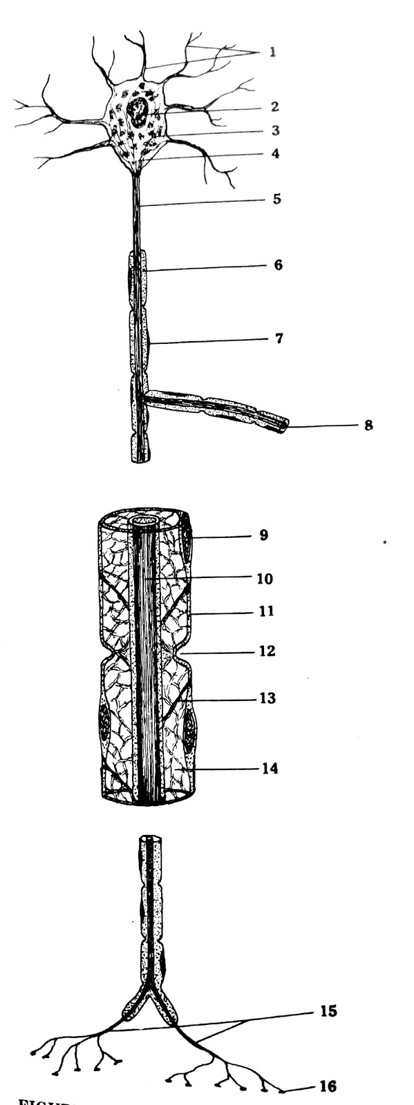
Fig:- Sehematic diagram of a typical neuron
- Dendrites
- Nucleus
- Nissl body
- Neurofibril
- Axis Cylinder
- Myelin sheath
- Nucleus of neurolemma
- Collateral brances
- Nucleus of neurolemma
- Axis Cylinder
- Neurolemma
- Node of ranvier
- Myelin incision
- Myelin sheath
- Terminal branches
- Terminal plate
Component parts of
the nervous tissue
Structurally,
nerve tissue consists of two types of cells
A. Nerve cells, or
neurons
B. Supporting
cells or glial cells
v Neuroglial cells and ependymal (CNS)
v Schwann cells and satellite cells (PNS)
A. Neurons
The structural and
functional unit in both the CNS and PNS is the neuron or nerve cell.
Composition
A neuron consists
of
i)
A
cell body, or perikaryon, and
ii)
Cell processes:-
Axon Dendrites
i) A cell body
Most neurons cell
bodies are situated in the grey matter of the brain and spinal cord.
Size: 5-100m
Shape: may be
pyramidal, fusiform(Spindle shape), flask shaped or polygonal
 Fig:-Multipolar Neurons, Spinal cord, sheep

- Axon hillock
- Dendrite
- Nissl granules
Features of cell body
a) Cell membrane
It is trilaminar
membrane: outer and inner protein layer, and intermediate lipid layer.
b) Nucleus
It is large,
spherical and central in position with a prominent nucleolus. Some nerve cells
may be binucleated.
c) Cytoplasm
It contains the
following structures;
Nissl bodies- Aggregation
of R.E.R & free ribosome (Protein synthesis)
Neurofibrils- Neurofilament
(Stability & support to the cell)
Microtubules
Mitochondria
Golgi-complex
Lysosomes
Centrosomes and
centrioles
Cytoplasmic
inclusions
ii) Cell processes
Axon
An axon is a
single long cylindrical process of neuron that varies in length and diameter
according to the type of neuron.
Features
- Axons are usually very long process for example, axons of the motor cells of the spinal cord that innervate the foot muscles may have a length of upto 100cm.
- Most neurons have only one axon.
- The axon arises from the cell body at the axon-hillock which is pyramid-shaped region.
- The plasma membrane of axon is called axolemma and its contents are known as axoplasm.

- Axon hillock
- Nissl granules
- Nucleus
- Dendrite
- Nucleolus
Dendrites
One or more, short
branching processes of neuron, specialized in receiving stimuli from the
environment, sensory epithelial cells, or other neurons are called dendrites.
Features
- One or more in number, each of which arise from the cell body
- They are usually short and divide like the branches of a tree
- Their cytoplasm contains Nissl bodies, mitochondria and neurofibrils
Function
Convey impulses
from periphery to the cell body. They are the principal signal reception and
processing sites on neurons.
Classification
of neurons
1. According to the number of cell
process, it is of three types;
i) Unipolar neurons
They have only one
process arises from one pole of the cell which divides into two branches one
which conducts impulses from sensory ending towards the cell body and another
which conveys these impulses to the brain or spinal cord. It is also known as
pseudo unipolar neurons.
Location:- Spinal
ganglia
Ganglia of certain cranial nerve
 Fig:- Dorsal root ganglion, spinal cord, chicken

- Axon
- Unipolar neuron
ii) Bipolar neurons
They have two
processes arises from two opposite poles of the cell. These processes are axon
and dendrite.
Location:- Ganglia
of inner ear
- Retina of eye
-Olfactory mucous membrane
iii) Multipolar neurons
Which have one
axon and two or many dendrites arises from different poles of the cell.
Location:- Neuron
of brain
-Spinal Cord
-Autonomic ganglia
2. According to the functional role
i) Motor neuron (efferent)
It carries
impulses from the CNS to the periphery which control effector organs such as
muscle fibre or glands.
ii) Sensory neuron (afferent)
It involved in the
reception of sensory stimuli from the environment and from within the body.
They conduct the impulses to the CNS for processing.
B. Supporting
cells or glial cells
Glial cells (Neuroglial cells) and ependyma (CNS)
Glial cells (Neuroglial cells) and ependyma (CNS)
Glial cells are
non-excitable supporting cells of the CNS, furnishing a micro-environment ideal
for neuronal activity.
Glial cells:- 3 types
Fibrous
Protoplasmic
b. Oligodendrocyte
2. Microglia
a. Astrocytes: 2 types
- Shape- star shaped
- Cell body- small (but largest of the 4 types)
- Cell processes- have a number of radiating processes
- Nucleus- oval
Fibrous:- Few long processes
Distribution:- White matter in the CNS
Protoplasmic:- Many short branched processes
Distribution:- White matter in the CNS
Protoplasmic:- Many short branched processes
Distribution:- Gray matter in the CNS

- Astrocyte, cell body
- Astrocyte, process
- Neuron
b. Oligodendrocyte:
- Smaller than astrocytes
- They have small cell bodies with a few delicate processes
- Nucleus- rounded and condensed. No microfilaments in their cytoplasm
Distribution:-
Predominant glial cells in the white matter in the CNS.
2. Microglia:
- Small elongated cells with short irregular processes
- Nucleus- dense elongated
- Somewhat less numerous than astrocytes and oligodendrocytes
- They originate from precursors cell in the bone marrow
Distribution:-
Both white and gray matter, usually near blood vessels
Acts as a phagocytic
cells in CNS, invade micro-organisms
3. Ependymal cells:
- Ependymal cells are low columnar or cuboidal cells that line the ventricles of the brain and central canal of the spinal cord
- In some CNS location, these cells are ciliated
- Tanycytes- special type of ependymal cell which extend processes into the hypothalamus

- Gray matter
- Central canal
- Cilia
v Responsible for secreting CSF
v Facilitate the movement of CSF
Schwann cells and satellite cells (PNS)
Schwann cells (Neuroloemmocytes)
- These are flattered cells
- Nucleus- ovoid or flattened
- Cytoplasm- Contains Small golgi complex and a few mitochondria
Function:- Allow
myelination of axon in PNS. One schwann cell forms myelin around a segment of one axon.
 Fig:- Nerve fascicles, unmyelinated , left ventricle, pig

- Perineurium
- Axon
- Schwann cell, nucleus
Satellite cells
- Small cells, derived from the embryonic neural crest like neurolemmocytes
- They form a covering layer over the large neuronal cell bodies in PNS ganglia
- Closely associated with the neurons and exert a supportive or tropic role

- Nerve fiber
- Neuron cell body
- Satellite cell
- Nisll granules
Myelin
sheath
Definition:
Myelin sheath is
lipoprotein complex that covers most of the axons and certain
dendrites in the central nervous system and PNS.
It is responsible
for the white matter of CNS and for white colour of many peripheral nerve.
Formation:
Myelin sheath is
formed by:
v Oligodendrocyte in the CNS, and
v Schwann cell in the PNS
Structure:
Myelin sheath
consists of many layers of modified cell membranes
Nodes of ranvier:
v Myelin sheath is interrupted at regular
intervals by gap, called the nodes of Ranvier, where the adjacent Schwann
cells/oligodendrocyte meet.
v Collateral branches of the axon arise at
the nodes of Ranvier.
v Nodes of Ranvier are the sites for the
exchange of ions between the axoplasm and extra cellular fluid.
Internode:
It is the distance
between two nodes of Ranvier and consists of one Schwann cell.
Functions: (Myelin sheath)
1. It acts as an
insulator of nerve fibre, and reduces the loss of electrical activities into
the surrounding tissue by dispersion
2. It is responsible
for the faster conduction of impulse through the nerve fibres. The impulse jump
from one node to another, the larger the intermodal segment, the faster is the
rate of conduction.
Myelin sheath is
absent in
i) Nodes of
Ranvier
ii) Proximal part
of axon (pre axon) close to cell body.
iii) Near the
termination of axon
iv) At the cleft
of Schmidt-Lanterman
Neurilemma (sheath of Schwann)
Surrounding the
myelin sheath, there is a thin membrane of Schwann cell that form a continuous
sheath and encloses the peripheral nerve whether myelinated or non-myelinated,
called neurilemma or sheath or Schwann.
v It is absent in the CNS
v In non-myelinated nerve fibre, the neurilemma
continuously surrounds axolemma.
Functions:
1. It helps in
protection and insulation.
2. It is
responsible for the regeneration of peripheral nerve fibres.
3. It is necessary
for the formation of myelin sheath (myelinogenesis)
Nerve fibres
A nerve fibre is
an axon (or dendrite) of a nerve cell with its covering, the function of which
is to conduct nerve impulses.
 Fig:- Dorsal root ganglion, dog

- Neuron cell body
- Nerve fibers
Classification
A.
(Structural basis)
1. Myelinated
fibres- Covered by myelin Sheath, and is white in colour.
Distribution:- White matter (CNS)
Peripheral nerves
 Fig:- Axons, myelinated, medulla, horse

- Myelin sheath
- Axon
Distribution:- Gray matter (CNS)
Post-ganglionic (ANS)
Somatic fibres (PNS)
 Fig:- Nerve fascicles, unmyelinated, uretar, pig

- Adipose tissue
- Nerve fibers x.s.
- Nerve fibers l.s.
B.
(Source of orgin)
1. Cranial nerves-
Arising from brain
2. Spinal nerves-
Arising from spinal cord.
C. (Functional basis)
1. Sensory
(Afferent) nerve fibres- carry sensory impulses from different parts of the
body to the CNS.
2. Motor
(efferent) nerve fibres- carry motor impulses from CNS to different parts of
the body.
D. (Distrubution basis)
1. Somatic nerve
fibres- these supply the skeletal muscles of the body
2. Visceral or
antonomic nerve fibres- these supply the various internal organs
E. (Chemical basis):- Neurotransmitter substance secreted
1. Adrenergic
nerve fibre- which secrete adrenalin
2. Cholinergic
nerve fibre- which secrete acetylcholine
(Myelinated
PNF) Structure:-
A myelinated
peripheral nerve fibre is made up of the following structures from within out
wards:
1. Axopalsm- forms
the central core of axis cylinder
2. Axolemma- semipermeable
membrane that covers the axoplasm
3. Myelin sheath- it
sorrounds the axis cylinder
4. Neurilemma- it
sorrounds the myelin sheath
Connective tissue covering
5. Endoneurium- it
is a delicate C.T. sheath which sorrounds the neurolemmal sheath. It
consists of a thin
layer of reticular fibres produced by Schwann cells.
6. Perineurium- it
is a C.T. sheath which sorrounds each bundles of nerve fibres. It is formed
by layer of flattened
epithelium like cells.
7. Epineurium- it
is the most external fibrous coat of dense C.T. which sorrounds the
bundles of nerve
fibres to form nerves
Nerve trunk
A collection of
bundles of nerve fibres(funiculi) enclosed in a connective tissue
sheath(epineurium) called nerve trunk. The main stem of a nerve fibres bound
together by a tough sheet of C.T.
Synapse
Synapses are the
sites of functional contact between neurons or between neurons and other
effector cells (eg; muscles and gland cells)
The synapse is
responsible for unidirectional transmission of nerve impulse from neuron to
another cell.
Classification
A) Anatomical (morphological)
1. Axosomatic synapse-
An axon of one neuron forms a synapse with a cell body of another neuron.
2. Axodendritic
synapse- Axon — dendrite
3. Axoaxonic- Axon
— Axon
4. Dendrodendrite
synapse- Dendrite — dendrite
B) Functionally
1. Excitatory
synapse
2. Inhibitory
synapse
C) Ultra structurally
1. Type—I
(asymmetrical synapse):
In which the
synaptic cleft is 30mm with pronounced thickening of the postsynaptic
membrane. Synaptic vesicles are spherical in this type.
2. Type—II
(Symmetrical synapse)
In which the
synaptic cleft is 20mm with apparent equal thickening of pre and post
synaptic membranes.
Synaptic vesicles
are flattened in this type.
Neurotransmitter is a chemical substance that act as the
mediator for the transmission of nerve impulses from one neuron to another
neuron through a synapse.
Neurotransmitters
are release at nerve endings and transmit impulses from nerve to nerve or from
nerve to effector tissue (muscle or glands)
If you have any questions you can ask me on :
mishravetanatomy@gmail.com
Twitter - @MishraVet
Facebook - Anjani Mishra
mishravetanatomy@gmail.com
Facebook - Anjani Mishra
Website: mishravetanatomy.blogspot.com
Post a Comment