
Epithelial Tissue
Written by Anjani Mishra

It's not clear who invented the first
microscope, but the Dutch spectacle maker Zacharias
Janssen (b.1585) is credited with making one of the earliest compound
microscopes (ones that used two lenses) around 1600.
The Dutch spectacle maker Hans Janssen and his son Zacharias are generally credited with creating these compound microscopes.
It was only until 1819 that Mayer coined the term “Histology”. He combined two Greek root words that are histos, for tissues, and logos, for study.
Due to the importance and originality of his contributions, the name of this new discipline devoted to the study of tissues was changed from 'tissue Anatomy' to 'Histology': this term had been coined in the early 19th century by Marie Francoise Xavier Bichat and reprised by Karl Mayer in 1819. The word Histology is derived from Greek word Histos + Logus/Logia where histos means tissu (French) or tissue (English) means weave/texture and logus/logia means logy (English) means study/knowledge. So, simply the meaning of Histology is the study/knowledge of tissue.
Definition
Histology is the
branch of Anatomy which deals with the microscopic form & structure (tissue) of an organisms.
Tissue
The world
"Tissue" is derived from the French word "Tissu" which
means weave or texture.
Tissue is an
association of cells with intercellular substance and fluids. The cells are
alike in form and function and are specialized for the performance of definite
task in the body.
There are 4 types
of basic tissues in the body :-
1.
Epithelial
tissue
2.
Connective
tissue
3.
Muscular
tissue
4.
Nervous
tissue
These tissues are
different from each other and one another structurally and functionally.
These differences
are due to differences in the physiological properties of these cells and other
composition with which the particular tissues are constituted.
Epithelial tissue

The word epithelium is derived from Greek word epi+thelion where epi means above and thelion means nipple. This tissue was
discovered at first by Marie Francois Xavier Bichat (French anatomist and pathologist) in between 1771-1802 in the nipple of mammary gland. So, the name has been given
as epithelial tissue. The Histologists described epithelium as a tissue which covers
the surface of the body as epidermis and lines all passages leading to the interior
(Eg; lining the wall of digestive, respiratory, and uro-genital system. etc.)

Functions:
- Providing protection- Skin
- Providing covering & lining
- Providing as mean for external & internal secretion
- Excretion- sweat, urine
- Absorption- intestinal mucosa absorb nutrients
- Sensation- tongue epithelium- taste bud and (Some epithelial cells get modified neuroepithelial cells)
Classification
On
the basis of lining/covering/membrane/surface
On
the basis of secretory glandular division
A.
On
the basis of Surface/lining division, it is of 3 types: namely;
1.
Simple
epithelium - tissue with single layer of cells
2.
Stratified
epithelium - tissue with more than one layer of cells
3.
Pseudostratified
- tissue with single layer of cells but appears to be stratified which is
actually a false
impression of stratification
(1) Simple
epithelium
One
the basis of morphology of cells it is of 3 types: namely;
- Simple squamous
- Simple cuboidal
- Simple columnar
(i) Simple squamous: it consists of single layer of cells. The
cells are flattened, fish scale like, plate like spindle shape, polygonal cell.
The nucleus is situated in the centre of a cell. The nucleus will follow the shape of the
cell (flat nucleus)
Lining :-
Lining :-
Endothelium -
Blood & Lymph vessels
|
Mesothelium -
Body cavity and Serous membrane
|
[It also lines the
parietal layer of Bowman's capsule, Visceral layer- modified sim. Sq. epi.
cells, called podocytes]

Fig: Simple squamous epithelium, mesothelium, liver, cat
- Squamous cell, nucleus
- Connective tissue
- Hepatocyte
(ii) Simple cuboidal: It consists of single layer of cells
which are equal in length and width. The nucleus is situated in the centre of a
cell and they are round in shape.
[Note: The term cuboidal is applied
when the height of each cell is approximately equivalent to its width.]
Lining the wall of smaller duct- Pancreas, kidney,
liver
- Surface lining of ovary.
- Thyroid gland (Follicles)
- Parotid and Mandibular salivary gland
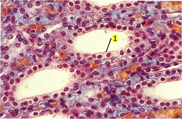
Fig: Simple cuboidal epithelium, kidney, cow
- Cuboidal cell
(iii) Simple columnar: It consists of single layer of cells. The
cells are higher in length than that of width. The nucleus is situated at the
centre or towards the basement membrane and they are oval in shape.
[Note: Nuclei are situated in the middle of the cells
when the cells are not secretory and at the base when they are secretory]
Ciliated-
Uterus, fallopian tube
|
Non-ciliated-
stomach (Glandular part), Intestine
|
 |
Fig: , Ciliated Simple columnar epithelium, infundibulum, oviduct, cow
|
 | |||
Fig: Non-ciliated Simple columnar epithelium, jejunum, dog |
- Columnar cell
- Lymphocyte
- Goblet cell
- Striated border
(2)
Stratified epithelium
Consist of more
than one layer of cells. On the basis of morphology of the surface cell layer,
it is classified into 4 types: namely;
i)
Stratified
squamous
ii)
Stratified
cuboidal
iii)
Stratified
columnar
iv)
Stratified
transitional
I.
Stratified Squamous
- Consists of more than one layer of cells (generally 3-5 layers, but it may be upto 30 layers)
- The basal layer consists of a single layer of columnar cells with oval nuclei
- The cells of middle layer are irregular, polyhedral is shape with round nuclei
- The cells of superficial layer are flattened with flat nuclei
It is of two types:
Keratinized
(dead layer of protein):- lining- skin, rumen, reticulum, omasum
Non-keratinized:-
lining- oesophagus, pharynx, vagina, mouth & nasal cavity
 | ||
Fig: Stratified squamous epithelium, keratinized, wattle, pig |
- Keratinized cells
- Dermis
 | |
Fig: Stratified squamous epithelium, non keratinized, esophagus, cat |
- Basal cell
- Esophagus, lumen
II.
Stratified Cuboidal
- Consists of more than one layer of cells
- The basal layer consists of a single layer of columnar cells
- The cells of superficial layer are cuboidal in shape with round nucleus located centrally
 | |
Fig: Bistratified cuboidal epithelium, esophagus, dog |
- Stratified cuboidal epithelium
- Smooth muscle
III.
Stratified Columnar
- Consists of more than one layer of cells
- The cells of deeper layer are usually cuboidal in shape
- The cells of middle layer are irregular, polyhedral is shape with round nuclei
- The cells of superficial layer are columnar in shape
nasal cavity(certain respiratory portion)
pharynx(certain portion)
anal canal(certain portion)
conjunctiva(certain parts)
 | |
| Fig: Stratified columnar epithelium, urethra, goat |
- Stratified columnar epithelium
IV.
Stratified Transitional
- Consists of more than one layer of cells
- The cells of basal layer is columnar in shape
- The cells of middle layer are irregular polyhedral shape
- The superficial cells are large, rounded/flattened in shape
 | |
| Fig: Stratified transitional epithelium, urinary bladder, cat |
- Transitional epithelium
(3) Pseudo-Stratified
epithelium
All the cells are located
on a common basement membrane with their nuclei at various level, but all the
cells are not reaching upto the surface. Nuclei of the cells are arranged into
many layers and this is due to the differences in the length of a cells and
this gives an impression of false stratification
It is of two types:
Ciliated
pseudostratified
Lining- Respiratory
tract (trachea, bronchi, bronchiole)
Non-ciliated
pseudostratified
Lining- Male
genital system (epididymis, ductus deferens)
 | |
| Fig: Ciliated pseudostratified columnar epithelium, trachea, cow |
- Columnar cell, ciliated
- Goblet cell
- Basal cell
- Basement membrane
A/to the number of cells present in the
gland, it is of 2 types;
i)
Unicellular-
it has only one cell, eg; goblet cell, unicellular salivary gland
ii)
Multicellular-
it has many cells, eg; liver, pancreas, testis, ovary
A/to the type/nature of secretion, glands
may be classified into 3 types;
i)
Serous
gland- secreting serous/watery fluid
Eg;
Sweat gland, Parotid Salivary gland
ii)
Mucous
gland- Secreting mucous substance
Eg;
Goblet cell
iii)
Sero-mucous
gland- Secreting watery fluid and mucous both
Eg;
Sub-lingual salivary gland, Mandibular salivary gland
A/to the mode of secretion, glands may be
classified into 3 types;
i)
Apocrine
gland (Apo means 'Apex', crine means to produce)
The
apex of a cell damage as a secretion. Eg; Sweat, mammary & uterine gland
ii)
Holocrine
gland (Holo means 'All')
The
entire cell damage as secretion eg; Testis, ovary, sabaceous gland
iii)
Merocrine
gland (Mero means 'part')
The
cells gives out their secretion without loosing any part. Eg; Liver, pancreas,
salivary gland
A/to the presence/absence of duct, glands
may be classified into 2 types;
i)
Exocrine
gland- glands with duct, eg; Sweat, salivary gland
ii)
Endocrine
gland- ductless gland, eg; Pituitary, adrenal, thyroid
A/to the duct system, glands may be classified
into 2 types;
i)
Simple
gland- with single duct system
Eg;
sweat gland, intestinal gland
ii)
Compound
gland- with branched duct system
Eg;
Mammary, Salivary gland
A/to
the size, shape and structure of the excretory unit of exocrine gland, it may
be classified into 3 types;
i)
Tubular
gland
Eg;
Intestinal gland
ii)
Alveolar
gland
Eg;
Sweat gland, pancreas
iii)
Tubulo-alveolar
gland
Eg;
Salivary glands
A/to
the morphology of the secretory unit of endocrine gland, it may be classified
into 3 types;
i)
Cord
type of gland
Eg;
Liver
ii)
Clump
type of gland
Eg;
Adrenal, pancreatic islet's
iii)
Follicular
type of gland
Eg;
Pituitary, Thyroid
Twitter - @MishraVet
Facebook - Anjani Mishra
If you have any questions you can ask me on :
mishravetanatomy@gmail.com Facebook - Anjani Mishra
Website: mishravetanatomy.blogspot.com
So nice
ReplyDeleteThis is very interesting and make easy to students to understand the epithelial tissue in brief .Thank you sir.
ReplyDeleteThank you so much sir. This is really helpful.
ReplyDeletemagnification ? Stains ?
ReplyDeleteThis is very helpful sir.
ReplyDeleteIt has greatly made veterinary study easier.
Useful 👏
ReplyDeletePost a Comment